Bunion Causes & Treatments

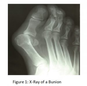
Bunions: Hallux Abducto Valgus is a deformity characterised by lateral deviation of the great toe, hence the most commonly used medical terms associated with a bunion anomaly are the terms ‘hallux valgus’ or ‘hallux abducto-valgus’ and ‘HAV’. The term hallux refers to the great toe or the ‘big toe’ and “valgus” refers to the abnormal angulation of the great toe which is commonly associated with bunion anomalies. This video shows the 3 stages of bunion deformity.


SAVE 10% OFF ICB PRODUCTS WITH DISCOUNT CODE: 02ICB10 Offer online only!

The use of the terminology abductus and abducto identifies that the direction of the hallux is away from the mid line of the body and approaches the second toe.

In the later stages of the bunion develop-mental progression the deformity can typically result in dislocation of the hallux from the metatarsal head1.

The answer to the question of ‘what causes a bunion’ is quite complex. A patient’s biomechanics is the main contributing factor behind the development of a bunion, however, tight fitting shoes can often aggravate the condition during its development.2

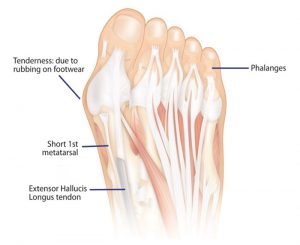
Excessive pronation will cause forces to be applied to the forefoot, with increased load on the 1st metatarsal head in an adducted direction. This will allow rotation of the shaft and in turn the hallux (big toe) will compensate by abducting.

A short 1st metatarsal or hyper mobile feet are considerably more susceptible, in this situation, the patient’s biomechanics is hereditary. As the 1st metatarsal adducts, rotates and drops to the ground to provide stability for the structure, a short 1st metatarsal shaft is a major contributing factor in the adduct-ing of the shaft to the midline of the body . When combined with pronation this caus-es the hallux to abduct, hence the term ‘Hallux Abducto Valgus’. (Lorimer et al, 1997; Selner et al, 1992; De Valentine, 1992).

In addition to the common HAV there is an alternate bunion known as a ‘Tailor’s bunion’, which is also described as a ‘bunionette’, a condition caused as a result of inflammation of the fifth metatarsal bone at the base of the 5th phalange or little toe.3

Similar foot mechanics issues appear to be the underlying causative factor being a shorter 5th metatarsal shaft which deviates or abducts away from the midline to gain ground control or stability , in turn the 5th digit adducts toward the midline of the body.

Three Stages of Bunion Development

Orthotic therapy at each stage can reduce the incident of the bunion progressing to the next stage.

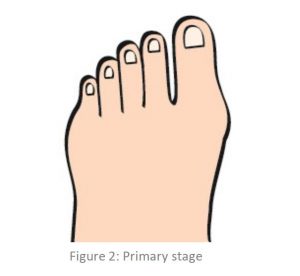
1.Primary Stage: usually occurs from adolescents up to the age of 25 years. A primary stage bunion presents as a slight bump See Fig 2

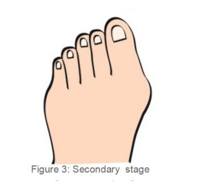
2. Secondary Stage; occurs generally at between the ages of 25 and 55 years. The 1st metatarsal head adducts and the hallux abducts causing pressure on the 2nd digit. Callosity may develop on the medial side of the 1st Metatarso Phalan-geal Joint and medial hallux. See fig 3

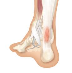
As the foot continues to pronate over several years the ground reaction forces en-courage the hallux into abduction and the extensor hallucis longus also becomes tight and pulls the hallux further across in a ‘bow’ like effect.

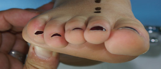
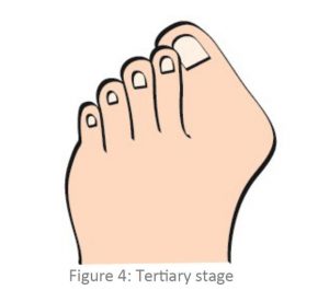
3. Tertiary Stage: If this situation is not controlled with orthotic therapy at the sec-ondary stage to prevent the condition from progressing further, the bunion will eventually move into the Tertiary Stage (or the 3rd stage– Fig 4). In the tertiary stage of Hallux Abducto Valgus an overlap-ping of the hallux occurs either above or below the 2nd digit. See fig 4 As this takes place the patient’s shoes become difficult to wear, and find it hard to find shoes to accommodate, as most shoes will aggravate the 1st MTPJ on the medial side (Thordarson, 2004).

This stage is very difficult to treat, the patient may be in extreme pain and often find it hard to find footwear that can accommodate for the deviated hallux. Patients may need to consult an orthopedic surgeon to surgically correct the bunion deformity. Following surgery, the patient will need to have orthotic prescribed to treat the underlying biomechanical condition. Orthotics are essential to give the foot realignment and support, and prevent the reoccurrence of the Hallux Abducto Valgus (bunion).

Treatment

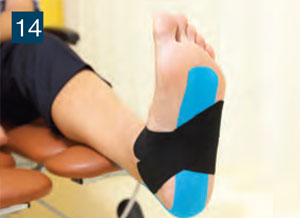
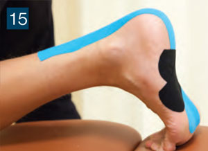
Night splints do not appear to be very successful in the correction of the HAV but may be of assistance to properly prescribed orthotic devices which will are designed to realign and control the patient’s excessive pronation thus reducing the development of the bunion. The night splint and alternatively bunion strapping may be of assistance during the first stage of bunion development, however the benefits may be undone when the patient engages in weight bearing activity without foundational correction

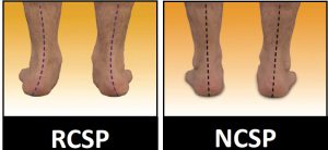
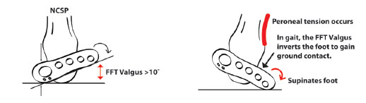
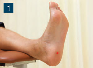
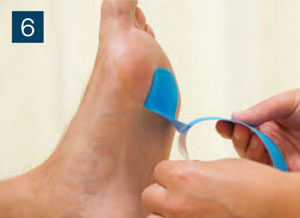
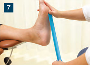
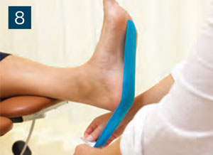
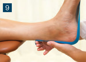
Prescribe an orthotic to realign and control the patient’s excessive pronation. Heat and mould the orthotic with the foot in the Neutral Calcaneal Stance Position (NCSP). Following heat moulding, monitor the patient, ensuring the orthotic is adequately controlling the excessive pronation – thus preventing further development of the bunion.

When treating hallux abducto valgus with orthotic therapy, it is important to explain not only the causes of bunion development but also the 3 stages. By doing so the patient will understand why they need to wear orthotics and that by doing so they will prevent the bunion from progressing to the next stage.

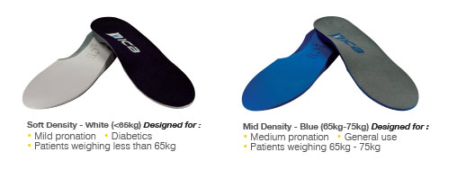
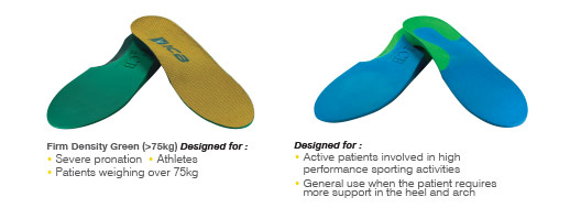
Treating Hallux Abducto Valgus with orthotic therapy will realign the foot, limit calcaneal eversion, thus controlling excessive pronation and taking pressure off the 1st MTPJ. However, monitor the bunion closely, if it worsens or continues to be painful, review the prescribed orthotic – make sure the orthotic is providing enough control, and check if the patient is continuing to excessively pronate through the orthotic. If the patient is continuing to excessively pronate it may be necessary to pre-scribe a firmer density orthotic such as the ICB Firm Green Orthotics, to ensure correction and control is being achieved.

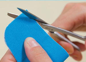
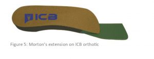
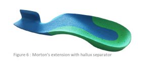
If the patient presents with a short 1st metatarsal shaft, the practitioner can create a Morton’s Extension on the orthotic. To do so place a Forefoot Orthotic Addition under the hallux (attach using double sided tape) – this treatment is only successful in the first stage.


References:
1. Thomas .C. Michaud Foot orthoses and other forms of conservative foot care 1997 P72
2.Bunions (Hallux Abducto Valgus)”. Footphysicians.com. 2009-12-18. Infor-mation Retrieved 2011-03-20- Tailor’s Bunion”.
General References
DE VALENTINE, S.J. (Ed) (1992) Foot and Ankle Disorders in Children, New York: Churchill Livingstone
LORIMER, D., FRENCH, GWEN, & WEST, S. (1997) Neales Common Foot Disorders: Diagno-sis and Management, 5th Edition, Melbourne: Churchill Livingstone
SELNER, A. J., SENER, M.D., TUCKER, R.A., & EIRICH, G. (1992) Tricorrectional Bunionecto-my for Surgical Repair of Juvenile Hallux Valgus, JAPMA
THORDARSON , DAVID B. (2004) Foot & Ankle, Lippincott Williams & Wilkins


Arch Pain When Wearing Orthotics

A common complaint amongst patient’s who have been prescribed foot orthotics is ‘pain in the arch’. This type of pain can be the result of 4 common issues:

1. Pain can be due calcification (similar to dupuytren’s contracture in the hand), a fibroma in the body of the Plantar Fascia, or a Ganglion cyst may be present.

TREATMENT: If there is calcification in the fascia, use manual therapy to break it down. For a fibroma or Ganglion cyst, a deflection will need to be heated into the orthotic to accommodate and relieve any pressure from this area.
Watch the video: Using heat to make a deflection in an ICB Orthotic.

2. Plantar fasciitis pain can be experienced at the attachment to the calcaneus.

TREATMENT: Control rearfoot pronation using orthotics with intrinsic rearfoot posting to realign the feet to the Subtalar Joint Neutral Position (STJN). If addi-tional inversion is required to control and achieve STJN, add extra rearfoot wedg-es (2° or 4°) to provide additional Calcaneal control1.

A medial arch infill can also be applied to the orthotic to provide increased arch support.

Watch the Video: Plantar fascial groove in a ICB orthotic. 

3. The Plantar fascia may be tight, and during gait (at mid stance to toe-off), compressing into the medial longitudinal arch of the orthotic causing discomfort and pain. To test for a tight fascia use the ‘Windlass Test’ see below.

Watch the video about the Plantar Fascial Groove.

TREATMENT: Create a plantar fascial ‘relief’ or ‘groove’ in the arch of the orthotic using heat or by grinding the orthotic. Place the groove 1cm from the medial edge through the arch contour.


4. The patient may exhibit unilateral excessive pronation as a possible compensation for a leg length discrepancy.

TREATMENT: Unilateral arch pain can be associated with a leg length difference2 due to long leg compensatory excessive pronation. If a structural leg length discrepancy is identified, a heel lift will need to applied to the orthotic on the shorter leg.

References:
1. FROWEN, P., O’DONNELL, M., LORIMER, D., BURROW, G. (2010) Neales Disorders of the Foot 8th Edition, p127 2. MICHAUD, T.C. (1997) Foot Orthoses and Other Forms of Conservative Foot Care, Sydney: William & Wilkins, p.114

When to Use Orthotics

One question that is often asked is ‘When should an orthotic be used in conjunction with the patients treatment program?

Many practitioners grapple with this issue and it can be confusing, especially if orthotic therapy is not used regularly in clinical practice.

One starting point is to examine the common conditions where orthotics are or have been recorded as being effective. The use of orthotics can be beneficial in many circumstances and so practitioners should be aware of the application and treatment. Doing this will make available to their patients treatment choices when attending to lower limb biomechanical conditions.

Practitioners should use their own modality in conjunction with orthotic therapy, or develop relationships with practitioners from other modalities, thus combining treatments to provide the patient with a holistic regime.
Orthotic therapy is not a definitive treat-ment, it should be regarded as just one part of the treatment protocol.

The common conditions that an orthotic can treat include:

  Bunions
usually caused by a short 1st metatarsal1 and aggravated by excessive pronation at  mid-stance to toe off 2.


  Ball of Foot Pain
collapsing and rotating of the metatarsals result-ing from excessive pronation.


  Plantar Fasciitis / Heel Spur
excessive pronation causes the fascia to elongate and tear from the calcaneal attachment. Spurs are a secondary compensation.


  Severs Disease
(children’s heel pain) – related to excessive pronation and growth spurts in children and affects sporting children more than sedentary ones.


  Achilles Pain
excessive pronation and supination creates a point of pain stress point.


  Shin Splints
lateral, medial, anterior – excessive pronation and supination are key contributing factors.


  Knee Pain
collateral ligament strain due to excessive pronation and supination factors.


  Osgood Schlatters Syndrome
occurs due to a combination of tibial tuberosity immaturity and quadriceps tightness3, growth spurts and tibial rotation factors.


  Hip Pain
due to structural or functional leg length and supination factors including tight external hip rotators.


  Low Back Pain
Unilateral and bilateral pronation, and structural and functional leg length causing stress on the lower back L1-L5.


  Leg Length Syndrome
when a structural leg length difference is evident the long leg may excessively pronate as compensation to level the pelvis.

A report by The American College of FOOT & ANKLE ORTHOPEDICS & MEDICINE entitled Prescription Custom Foot Orthoses – Practice Guidelines, December, 2006 names the following conditions as being treatable with orthotic therapy :

1. Proximal Lower Extremity Pathology A. Shin Splints B. Tendonitis (Tenosynovitis) C. Posterior Tibial Dysfunction D. Chondromalacia Patella (Runner’s Knee, Patellofemoral Syn-drome) E. Iliotibial Band Syndrome F. Limb Length Discrepancy

2. Arthritides A. Inflammatory Arthri-tis, B. Rheumatoid Arthritis, Psoriatic C. Arthritis, Other Inflamatory Ar-thritides D. Osteoarthritis

3. Mechanically Induced Pain and Deformities A. Pes Cavus, Haglund’s Deformity B. Hammer Digit Syndrome C. Functional Hallux Limitus, Hallux Limitus and Hallux Rigidus D. Plantar Fasciitis E. Equinus F. Sinus Tarsi Syn-drome G. Tailor’s Bunion (Bunionette) H. Hallux Abducto-Valgus (Hallux Val-gus, Bunion) I. Pes Planus J. Metatarsal-gia K. Sesamoiditis L. Morton’s Neuroma (Intermetatarsal Neuroma)

4. Pediatric Conditions A. Calcaneal Apophysitis B. Genu Varum and Genu Valgum C. Tarsal Coalition D. Metatarsus Adductus 5 . Sensory Neuropathies A. Peripheral Neuropathy B. Charcot Neuroarthropathy (Charcot Foot)C. Tarsal Tunnel Syndrome So what is the starting point? And when should I use orthotics? First ,establish if the patient presents with a condition that is recorded as, being able to be treated with orthotics.

Check for pronation or supination. If the patient pronates, this will often be an underlying factor to many of the conditions mentioned above, treatment with orthotics to realign and control the rearfoot and support the longitudinal arch should be undertaken.

Identifying Lower limb biomechanical anomalies and foot mechanics issues relating to both pronation and supination will be a key treatment to realigning and controlling the rearfoot with orthotics.

Identify the amount of excessive pronation or excessive supination by correcting the foot to neutral and then allowing the patient to rest and relax their feet (NCSP and RCSP)* this will allow you to identify whether the patient will require a corrective orthotic product.

Next, check structural leg length and if a heel lift is required attach to the orthotic for the short leg, start by using ½ the measured amount or a 4mm heel lift addition. The long leg may be excessively pronating as compensation and will need to be supported by the orthotic. Never use a single orthotic, always prescribe a pair as this will maintain correct foundational balance.

If the foot is supinating the patient will experience jarring in the foot and to the upper structure, and will often have a rigid high arched foot type. This type of foot commonly exhibits a forefoot valgus deformity. The pes cavus type foot is not that common – about 8 – 9% of the population may present with it.

In this case an orthotic is required to control the supination by maintaining the Ideal / Neutral Calcaneal Stance Position(NCSP), use a forefoot addition to treat the forefoot valgus, whilst providing the patient with comfortable support.

Prescribing an orthotic for your patient base is easy as: 1, 2, 3

1. Identify the RCSP (patients Resting position).

2. Observe the NCSP (Neutral or corrected position) to identify the correct position for heat moulding the orthotic, and to help identify the pronation effect,
i.e. NCSP – RCSP = Total Pronation
Observation of Rearfoot positioning and Anterior positioning using ICB Anterior Alignment Method ( ICBAAM) see youtube.com/icbmedical


3. Check the leg length (manually) for any structural differences.

4. The Palpation for Supine Medial Malleoli Asymmetry Technique is an easy method which has been reported as having both Intra-examiner and inter-examiner reliability. The technique was clinically trialled at RMIT University 2005 by Gary Fryer4.
By following these simple steps more than 80% of the Lower Limb Biomechanical conditions practitioners see in the clinic on a daily basis can be treated effectively with orthotic therapy.
Patient’s should be instructed to wear their orthotics for 1-2 hours per day, increasing gradually over 1-2 weeks until it is comfortable wearing them full time. If discomfort is experienced, the patient should take the orthotics out of their shoes, and give their feet a rest. Continue until it is comfortable wear-ing the orthotics all day.

“When should orthotic additions be attached to the orthotic?”
Orthotic additions can be added on the subsequent consultation.
Forefoot & Rearfoot additions can be attached to the orthotics on subsequent visits – if required.

If you need any further assistance or advice with patient assessment, or prescribing orthotics View DLT Facebook, contact DLT Podiatry
email: sales@dltpodiatry.co.uk, or go to the website: www.dltpodiatry.co.uk


ICB Orthotic demonstration

ICB Orthotics

I would like to arrange an ICB Orthotic demonstration at my clinic and claim my free pair of ICB Orthotics.

The demonstration will include heat moulding and how to place the foot into the neutral position and take no longer than 30 minutes.

    ICB Course Dates 2018

    ICB Superior BiomechanicsICB Courses 2018


    This course will present a foundational understanding of lower limb biomechanics by independent pre-learning together with a workshop on practical orthotic therapy. Taught by Lawrence Dreifuss owner of Tuckton Chiropody & Podiatry Centre.

    Book Now

    PRACTICAL HANDS ON WORKSHOP OBJECTIVES:

    This practical ‘hands on’ day will provide the manual skills to implement chairside orthotic therapy to fulfil a successful treatment plan for your patient.
    Upon completion of this workshop, the participants will be able to:
    ▪ Identify Neutral Calcaneal Stance Position (NCSP)
    ▪ Assess for Structural Leg Length Discrepancy
    ▪ Heat and moulding orthotics to the patient’s STJN

    Evaluate and treat forefoot mechanics, including:
    ▪ Forefoot Valgus
    ▪ Forefoot Varus
    ▪ Plantarflexed 1st
    ▪ Dorsiflexed 1st

    Perform chairside orthotic modifications, including:
    ▪ Plantar fascial grove to treat tight Fascia
    ▪ Extrinsic and Intrinsic metatarsal Dome for Metatarsalgia and Mortons Neuroma
    ▪ 1st Metatarsal deflection to treat plantarflexed 1st
    ▪ Forefoot Varus and Forefoot Valgus posting
    ▪ Morton’s extension for HAV, Dorsiflexed 1st & Hallux limitus.

    Attendees will receive a starter pack (worth over £100) which will include a pair of orthotics to heat mould, plus a set of products to deflect and customise in the workshop.

    We provide prior learning 30 days before the course, to ensure all participants benefit from the subjects taught on the day.

    Book Now

    Dates are:

    OR CONTACT RICHARD ON 07553 345 831OR EMAIL richard@dltpodiatry.co.uk


    FULL !! FULL!!! SUNDAY 4TH MARCH
    1 Day Course: £90 + VAT
    INCLUDES ICB Starter Kit worth over £100 + VAT

    AT DLT PODIATRY SUPPLIES – HUDDERSFIELD- HD1 4TW
    Riverside Court, Stoney Battery Road, Huddersfield, West Yorkshire
    BOOK


    OPEN DAY

    Free entry and open to all, special offers and discounts available on the day. We bring our showroom to you, view new products and equipment. See product demonstrations by our friendly team who are happy to give help and advice.

    FULL !! FULL!!!  SUNDAY 18TH MARCH
    1 Day Course: £90 + VAT
    INCLUDES ICB Starter Kit worth over £100 + VAT

    Doubletree by Hilton NORTH – NEWBURY – RG20 8XY
    M4, Junction 13, Oxford Road, Newbury RG20 8XY

    BOOK

    OPEN DAY

    Free entry and open to all, special offers and discounts available on the day. We bring our showroom to you, view new products and equipment. See product demonstrations by our friendly team who are happy to give help and advice.


    Saturday 15th September – Heathrow
    1 Day Course: £110 + VAT
    INCLUDES ICB Starter Kit worth over £100 + VAT

    Premier Inn , Bath Rd, 15 Bath Rd, Heathrow, Longford, Hounslow TW6 2AB

    BOOK


    13th October Manchester  

    Saturday 13th October – Manchester
    1 Day Course: £110 + VAT
    INCLUDES ICB Starter Kit worth over £100 + VAT

    Premier Inn Trafford Centre West – Old Park Ln, Stretford, Manchester M17 8PG

    BOOK

     TO BOOK ONLINE CLICK HERE


    BIOGRAPHY: LAWRENCE DREIFUSS

    Lawrence Dreifuss

    After graduating from the London Foot Hospital
    in 1987, Lawrence worked within NHS Community and hospital clinics before establishing Tuckton Chiropody & Podiatry Centre in 1997 with his Podiatrist wife Sheila.

    Expansion into a multi chair clinic and the recruiting of associates followed allowing Lawrence to specialise and further hone his knowledge & interest of Podiatric Biomechanics Tuckton Podiatry now boasts its’ own separate Biomechanical suite adjoining the main clinic, utilising up to the minute  technology and techniques, treating patients with the skeletal / muscle issues patients face as a result of their foot dynamics or trauma.

    Lawrence specialises in treating musculo-skeletal conditions of the lower limb and postural abnormalities related to gait / pelvic dysfunction especially related to chronic sports injury.

    Inversion Sprain & the Effects of Forefoot Valgus

    FREE POSTER

    This article will focus on lateral sprain and pain associated with a pes cavus foot structure and a forefoot valgus deformity. Repetitive lateral ankle sprain or lateral knee pain (or even lateral shin splints) is often diagnosed as ‘idiopathic’, closer examination of the biomechanical relevance needs to be pursued. The term ‘idiopathic’ is often used in this area as there seems no reason for the pain occurrence. Pes cavus foot (high arch) structures (Fig. 2) may have a predisposition to lateral ankle sprains and present as a rigid structure and a supinated foot structure.


    Pes Cavus Foot Structure

    FIG2

    This type of structure will usually exhibit a forefoot valgus deformity
    meaning that, ‘the plantar plane of the forefoot remains everted relative to the plantar plane of the rearfoot when the subtalar joint is in neutral.’


    This deformity will have an impact on the patient in heel strike, midstance and toe-off phases of gait.  The patient who exhibits a pes cavus foot structure will often present with a forefoot valgus (FFVL) greater than 10º and also often exhibit a plantar flexed 1st metatarsal

    (Boyd & Bogdan, 1993) – encouraging the foot to strike laterally and eliciting pressure on the lateral aspect of the hip joint.  If the forefoot valgus deformity is greater than 10º, the foot will often continue to supinate through the cycle, having a ‘jarring’ effect on the upper structure, putting additional strain on the lateral aspect.

    Fig. 3

    Fig. 3: Use an ICB Protractor to assist in
    measurement of Forefoot valgus.


    When the foot is supinated it often exerts stress on the peroneals and may cause elongation of the muscles and tendons, thus weakening the retinaculum and lengthening the peroneals, often causing the tendon to sublux off the lateral aspect of the malleolar.

    The forefoot valgus deformity (in gait) encourages the foot to invert the foot, propulsion is delayed causing lateral instability and results in tension and tearing of the peroneal muscles, causing inflammation and tenderness, and difficulty walking. Lateral ankle sprains are more common than medial due to the fact that ligaments are weaker on the lateral side.

    Hence the lack of lateral stability can be caused by uncompensated or partially compensated rearfoot, a flexible forefoot valgus or osseous forefoot valgus (Boyd & Bogdan, 1993; Hollis et al, 1995; Shapiro et al 1994).

    Fig 4

    Fig. 4: Effects of Forefoot valgus during gait.

    There are also certain biomechanical foot deformities that make some patients more susceptible to inversion sprains, such as, neurological deficits and supinated foot types which exhibit or function with a supinated calcaneus (Valmassy, 1996).


    In summary, if a patient presents with lateral hip pain, knee pain, ankle strain or repetitive lateral inversion sprain, always check for a forefoot valgus deformity.  If a forefoot valgus if present, add an appropriate size ICB Forefoot Valgus wedge (available in 4° & 6°) to the selected ICB Orthotics using the 3M tape provided.  Next, heat mould the ICB Orthotics to the patient’s Neutral Calcaneal Stance Position. Being made from 100% EVA, ICB Orthotics can easily be heated and moulded to suit high arch foot structures.

    ICB Orthotics

    Prescribing ICB Orthotics.

    ICB Heat Moulding Orthotics can be prescribed to assist in the treatment of excessive pronation and supination, and resultant biomechanical conditions.  With 5 densities, 5 styles & 11 sizes, there is an ICB Orthotic to suit all footwear styles, patient ages and activity levels.

    ICB Orthotics

    ICB OrthoticsPRICES START FROM £18.85

    To enable quick and easy orthotic customisation in the clinic setting, ICB Orthotic Additions are also available, including: Forefoot & Rearfoot wedges, Heel Lifts, Metatarsal domes, medial flanges,
    medial arch infills and more.

    View the full ICB Orthotic range


    Dr.’s Remedy Top 10 Best Sellers

    Dr.'s Remedy Top Sellers

    Dr.’s REMEDY podiatrist formulated nail care collection has proved popular amongst Podiatrists and Health Care practitioners due to the addition of Tea Tree and the fact that you can use with topical fungus medications which will not make the condition worse.

    With 37 colours now in the collection we have accumulated the Top 10 sellers in the past 12 months.

    Dr.'s Remedy Mindful Mulberry Dr.'s REMEDY Peaceful Pink Coral

    No.1: Dr.’s REMEDY Mindful Mulberry
    REM39 –
    This renewed juicy berry shade is stylishly tart and playful yet sweet and classic.
    A perfect feminine neutral. VIEW

    No.2: Dr.’s REMEDY Peaceful Pink Coral
    REM16 –
    A poppy, fresh bubble-gum shade   VIEW

     Dr.'s REMEDY Positive Pastel Pink Dr.'s REMEDY Cozy Cafe

    No.3: Dr.’s REMEDY Positive Pastel Pink
    REM17 –
    Baby girl pink with swirls of
    pale purple.
    VIEW

    No.4: Dr.’s REMEDY Cozy Cafe
    REM6 –
    A cafe au lait with hints of pinkish grey and a splash of lilac.
    VIEW
    Dr.'s REMEDY Focus Fuchsia Dr.'s REMEDY Playful Pink

    No.5: Dr.’s REMEDY Focus Fuchsia
    REM8 –
    A hot pink classic with rich, romantic allure.
    VIEW

    No.6: Dr.’s REMEDY Playful Pink
    REM34 –
    A cheerful candy pink with a subtle flamingo pink –
    VIEW

    Dr.'s REMEDY Relaxing Rose Dr.'s REMEDY Clarity Coral

    No.7: Dr.’s REMEDY Relaxing Rose
    REM20 –
    An opaque, vintage rose petal shade
    with a hint of iridescence.
    VIEW

    No.8: Dr.’s REMEDY Clarity Coral
    REM5 –
    Bright pinky orangey and matte.
    VIEW

    Dr.'s REMEDY Remedy Red Dr.'s REMEDY Balance Brick Red

    No.9: Dr.’s REMEDY Remedy Red –
    REM21 –
    A true iconic old-Hollywood red.
    VIEW

    No.10: Dr.’s REMEDY Balance Brick Red –
    REM3 – A deep modern maroon.
    VIEW

     VIEW ALL REMEDY COLOURS


    INTERESTING FOOT FACTS…………

    16473482_1610921962257087_2699836974541235257_n

    Management of Achilles tendinopathy in runners-LER 
    By Howard Kashefsky, DPM
    Achilles tendinopathy is a common lower extremity injury in athletes as well as nonathletes. The Achilles tendon is often a site of injury in runners and is the second-most common running-related musculo¬skeletal injury, after medial tibial stress syndrome, with an incidence of 9.1% to 10.9%.
    1 The lifetime risk in former elite male distance runners is 52%.
    2 Factors that may contribute to Achilles tendinopathy include overuse, systemic disease, older age, sex, body composition, and biomechanics.
    3 Elevated biomechanical load has been shown to cause both microscopic and macroscopic failures.4-6


    1. Lopes AD, Hespanhol Junior LC, Yeung SS, Costa LO. What are the main running-related musculoskeletal injuries? A systematic review. Sports Med 2012;42(10):891-905.
    2. Zafar MS, Mahmood A, Maffulli N. Basic science and clinical aspects of Achilles tendinopathy. Sports Med Arthrosc 2009;17(3):190-197.
    3. Magnan B, Bondi M, Pierantoni S, Samaila E. The pathogenesis of Achilles tendinopathy: a systematic review. Foot Ankle Surg 2014;20(3):154-159.
    4. O’Brien M. Functional anatomy and physiology of tendons. Clin Sports Med 1992;11(3):505-520.

    VIEW ICB ORTHOTICS


    COURTIN Total Footcare

    Courtin Total Footcare

    Why Courtin?

    A skincare line based on 100% pure Tea Tree oil. Excellent treatment to help many skin problems. Compact and complete series of products.
    Courtin has developed a special total skin care and foot care line containing Original Australian Tea Tree oil. The Courtin Tea Tree range includes a wide variety of salon and direct sale products.

    What is Tea Tree oil?
    Tea Tree oil is produced from the leaves of the Melaleuca Alternifolia by means of a technological process using steam. This process is subject to painstaking quality controls. The result is a purity level of 100%. Since it is a pure and natural product, the colour of the pure oil product can vary from colourless to light yellow.

    Tea Tree oil can stimulate the healing of the conditions listed below
    • Bacteria, fungi and viruses
    • Ingrown nails
    • Itching
    • Cracked/chapped skin
    • Corns
    • Inflamed cuticles
    • Psoriasis
    • Chilblains: toes, feet or hands
    • Warts
    • Excessive sweating of feet
    • Athlete’s foot

    Pure Tea Tree Oil

    Ways to use Tea Tree Oil 

    Nail Infections – Apply a few drops of Tea Tree oil to the infected area twice per day and allow to penetrate. Can be used on both hands and feet. Place the hands or feet in warm water bath containing approximately 10ml of Courtin Foot Bath and add 1-2 drops of Pure Tea Tree Oil to the bath. After bathing, pat the hands and feet dry and massage with Courtin Hand & Foot Cream. Retreat the infected areas with Pure Tea Tree Oil.

    Sprains – Rub Pure Tea Tree Oil directly on the sprain to ease the pain. Thoroughly wash and dry the feet. Apply Courtin Hand & Foot Cream and massage until absorbed. Apply Pure Tea Tree Oil to the affected areas twice per day.

    Perspiring feet – Add 5-10 drops of Tea Tree oil to warm water. Take 5 minute foot bath every evening with Courtin Foot Bath. After the foot bath, apply Courtin Hand & Foot Cream and massage until absorbed. Put several drops of Tea Tree Oil in your shoes or apply some Courtin Foot Deo Spray.

    General applications for various conditions:

    Vaporisation
    To purify the air, place a few drops on a handkerchief, sheet or cloth placed on a radiator or in the hanging radiator humidifier.

    Foot Bathing
    2 – 3 drops of Tea Tree oil in the bath water combined with the Courtin Footbath.

    Compresses
    Add 2 – 4 drops of Pure Tea Tree Oil to 10 cl. of water mixed with Courtin Footbath. Mix well. Soak the compresses in the Tea Tree oil solution and place them on the site to be treated for several minutes; repeat several times. Adjust the temperature of the water to the ailment being treated.

    Massage
    For massages, Tea Tree oil must always be mixed with:
    ■ For the Feet – Courtin Hand & Foot Cream.

    When massaging the face, do not exceed a maximum concentration of 3% essential


    Courtin treatments with your pedicure
    Your pedicure offers various treatments with the wide range of Courtin Tea Tree products. The Courtin treatments not only act preventively but are exceptionally effective for many foot conditions.

    Spa Foot Mask

    Cosmetic Spa Foot Care Treatment – For most of the day, our feet are enclosed in footwear. This treatment with the unique Spa Foot Mask is a real blessing and you can actually feel the energy flowing back into your feet. A cosmetic and nourishing foot treatment providing total relaxation for body and soul.

    Foot Mask – For some extra pampering, you can treat your feet with a nourishing foot mask. After a foot peeling, the toenails and cuticles are massaged with nail oil. After that, a foot mask is applied followed by a massage with a specially formulated, nourishing foot cream.

    Courtin Foot Care Home Products

    Pure Tea Tree Oil

    100% Pure Tea Tree Oil – The 100% PureTea Tree Oil is made using natural processes and without additives. It is extremely versatile to use and has cell-renewing, pain relieving and soothing effects.

    Courtin Hand & Foot Cream

    Hand & Foot Cream  – This Hand & Foot Cream both cleanses and soothes. In addition to it’s cleansing and soothing properties, it also prevents odours. After cleansing, apply to dry feet and massage until absorbed.

    COURTIN Chapped Skin Cream

    Chapped skin Cream – The perfect cream for feet with highly chapped soles which can lead to cracks and fissures of the soles. With regular use, this cream softens the chapped skin, helps to prevent cracking and stimulates the healing of your feet. also ideal for raw skin on top of the feet. After cleansing apply to dry feet and massage until absorbed.


    Courtin Foot Bath

    Foot Bath – For intensive cleansing and reinvigoration of tired feet, use Courtin Foot Bath. Your feet will feel smooth and fresh again. Prevents unpleasant odours. Mix a spoonful of the gel into approximately 4 litres of water. Bathe the feet in water for 5 min.


    Courtin Nail Oil

    Nail Oil – Contains extremely nourishing active ingredients. Returns a natural shine to the nails. Apply once per day and massage for several minutes until the oil has been absorbed by the nail.

    Courtin Ice Gel

    Ice Gel– For the invigoration of tired, heavily stressed and burning feet or legs. The Ice Gel is directly absorbed through the skin. It prevents unpleasant odours and stimulates the circulation. Massage from the sole of the foot upward toward the thigh.

    Courtin Foot Deo Spray

    Foot Deo Spray – The Foot deodorant Spray keeps the feet fresh and any unpleasant odours are neutralised. Spray the Foot Deo on the tops and soles of the feet and rub it in. Don’t forget to apply between the toes.


    Hapla Tape For Plantar Fasciitis

    hapla

    Before applying tape it is important
    to remember to round the corners
    of each piece.
    Rounding the corners prevents the
    tape from catching on clothes and
    peeling back which may reduce the
    time the tape will stay adhered to the
    patient.
    How to cut hapla tape
    Directions for use – Rounding Corners
    1 Plantar Fasciitis - Hapla Wave Tape 2 Plantar Fasciitis - Hapla Wave Tape

    Patient will be experiencing pain either on the central or medial portion of the heel. Ensure the foot is in a dorsiflexed position.

    Cut a full width piece of tape long enough to go from the metatarsal heads up to the mid calf.

     3 Plantar Fasciitis - Hapla Wave Tape 4 Plantar Fasciitis - Hapla Wave Tape

    Tear and remove the backing paper from one end of the tape.

    Place an anchor point at the metatarsal heads applying zero stretch.

    5 Plantar Fasciitis - Hapla Wave Tape 6 Plantar Fasciitis - Hapla Wave Tape

    Remove the first part of the remaining backing paper.

    Apply 50% stretch to the tape.

    7 Plantar Fasciitis - Hapla Wave Tape 8 Plantar Fasciitis - Hapla Wave Tape

    Apply the tape down to the heel.

    At the heel reduce the stretch down to 25%.

    9 Plantar Fasciitis - Hapla Wave Tape 10 Plantar Fasciitis - Hapla Wave Tape

    Apply tape to the Achilles.

    Place an anchor point with zero stretch on the achilles tendon and apply friction to all areas of the applied tape.

    11 Plantar Fasciitis - Hapla Wave Tape 12 Plantar Fasciitis - Hapla Wave Tape

    Cut a six inch length of tape, tear and remove the central part of the backing
    paper.

    Apply a 50% stretch across the site of pain. Make sure you apply each end (anchor points) with zero stretch.

    13 Plantar Fasciitis - Hapla Wave Tape 14 Plantar Fasciitis - Hapla Wave Tape

    Apply another six inch length of tape at a slightly more oblique angle to the first
    across the site of the pain.

    Apply friction to all areas of the applied tape and ensure patient is comfortable.

    15 Plantar Fasciitis - Hapla Wave Tape

    Alternatively, if they are able, you could put your patient into this position to ensure dorsiflexion of the foot.