Monthly Archives: March 2019

Osgood–Schlatter Disease

ICB Lower limb biomechanics

Osgood–Schlatter disease or syndrome, also known as Apophysitis of the Tibial Tubercle, is an irritation of the patellar ligament at the tibial tuberosity occurring at the tendon-bone junction of the patellar tendon and the tibial tuberosity.1

The condition is named after Robert Bayley Osgood (1873-1956), an American Orthopedic surgeon and Carl B. Schlatter, (1864-1934), a Swiss surgeon who described the condition independently in 1903.

Watch the video below about Osgood–Schlatter disease below.


View ICB Products

General medical opinion is that Osgood Schlatter Disease, is caused by repetitive stress or tension on the growth plate of the upper tibia (the apophysis), which can be complicated by growth spurts and biomechanical deformities or anomalies. In general medical circles it is often called ‘growing pains’ and is similar in bio-mechanical dysfunction to Severs Condition, however occurring in this instant at the tibial tuberosity being characterised by inflammation of the patella tendon and surrounding soft tissues at the point where the tendon attaches to the tibia.

Osgood Schlatter Disease

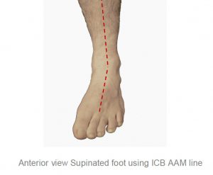
This condition appears to afflict children who are growing fast and often have external Tibial Torsion condition combined with Internal Tibial Rotation associated with excessive pronation.

Osgood-Schlatter’s Disease, is we believe, caused by repetitive stress or tension on the growth plate of the upper tibia (the apophysis), which can be complicated by growth spurt syndrome and biomechanical deformities or anomalies. The cause is similar in biomechanical operation to Sever’s Disease, except that it occurs at the tibial tuberosity and characterised by inflammation of the patella tendon and surrounding soft tissues at the point where the tendon attaches to the tibia.

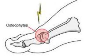
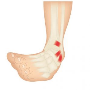
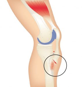
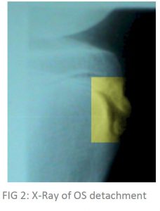
As stated, it is usually young people who suffer with this disease – experiencing pain just below the knee joint and patella which usually worsens with activity. It is also associated with an avulsion injury (stretching the tendon so much that it tears away from the tibia and in extreme cases takes a fragment of bone with it – See Figure 2).

Osgood Schlatter’s Condition

A bony bump may appear on the up-per edge of the tibia (below the knee cap) that may be particularly painful when external pressure is applied. It has been misdiagnosed in the past in Australia as “surfer’s knee” (a myth that only surfboard riders suffered from the condition). The hinge motion of the knee is not actually affected.

Most commonly Osgood Schlatters Disease affects active young people, particularly boys between the ages of 10 and 16 who play games or sports that include frequent running and jumping.

In a retrospective study of adolescent athletes actively participating in sports showed a frequency of 21% reporting the syndrome compared with only 4.5% of age-matched non-athletic controls2. Bilateral symptoms are observed in 20–30% of patients indicating that there is a higher incident of unilateral occurrence lead-ing one to deduce that in the unilateral cases, structural leg length or other unilateral biomechanical anomalies may be a contributing factor.

View ICB Products

Symptoms of Osgood Schlatters Disease include:

• Pain over the tibial tubercle
• Swelling over the tibial tubercle
• Weakness in the quad muscle group
• Increased pain & swelling with activity
• A visible lump at the base of the knee cap
• Pain to the touch over the affected area.

Often the pain may last only a few months or may recur until the child stops growing.

Not a lot has been written about the effect of the combination of a growth spurt combined with both pronation or supination, and internal and external tibial torsion. However, children appear to experience more pain and resultant damage to the attachment when these factors occur in combination.

The pain (and resultant effects) become more noticeable during activities that require running, jumping or going up or down stairs and is most common in young athletes who play football, soccer, basketball, netball or who are involved in gymnastics and ballet.

Contributing Factors whilst the Child is still growing

1. Excessive Pronation:
As the foot pronates internal tibial rotation occurs. The body’s mechanism of compensation causes the ITB’s and abductor muscles to tighten in the opposite direction causing the patella to laterally and superiorly displace, hence causing a tractional pull on the tibial tuberosity.

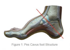
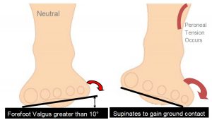
2. Supination & Pronation:

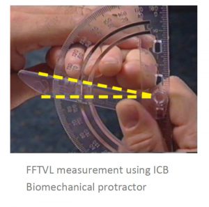
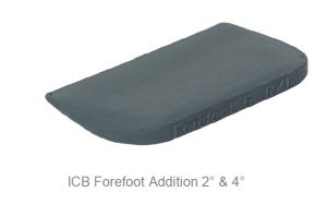
Because of the anterior position of the tibial tuberosity, when the foot supinates due to a FFT valgus deformity less than 10 degrees in the swing phase of gait, the foot lands laterally then the ground reaction forces (on the lateral side) propel the foot into pronation with the same effect above.

3. Internal Tibial Torsion:

Internal tibial torsion will cause the foot to rotate the tibial condyle medially. The ITB’s and gluteals then tighten and come into action as an external rotator of the femur as the compensatory mechanism causing external rotation above the knee and internal rotation below the knee.

The two rotation actions have an effect on the patella and its tendon attachment also causing the tibial tuberosity to stretch and pull.

4. External Tibial Torsion: This causes the foot to externally rotate in the swing phase of gait, the iliopsoas and adductor muscle groups combined to provide compensation at the late swing phase, causing the foot to straighten and land laterally (same effect as 2). Then the ground reaction forces cause the foot to pronate with the help of the psoas pull resulting again in internal rotation above the knee and external tibial torsion position increases tractional forces on the patella and its attachment.

Treatment

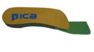
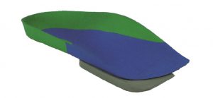
• Orthotics moulded to the patient’s Ideal/Neutral Calcaneal Stance Position to realign and hence reduce the effect of tibial rotation.

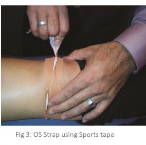
• Combine orthotic therapy with strapping: use an Osgood Schlatters strap (see below) to reduce the tension on the attachment at the growth plate whilst the child is playing sport (see Figure 3).

Strap Using Sports Tape

Stretching may exacerbate the tearing – alternatively use deep friction massage to help in pain relief.

• R.I.C.E
Active children may experience shortening of the muscles whilst growing, which coupled with biomechanical anomalies = predisposition to Osgood Schlatter.

Contraindications
It is important to differentiate from malignancy, infection, fracture, tendonitis and Sindling-Larsen-Johansson Disease. Initially the diagnosis is based on clinical signs and symptoms including: pain, heat, tenderness and local swelling with prominence at the tibial tuberosity. X-ray is required to establish the extent of the condition.

Steroid injections are discouraged as this may cause weakening of the infra-patellar ligament, scaring and fat necrosis.

View ICB Products

REFERENCES
1 Nowinski RJ, Mehlman CT (1998). “Hyphenated history: Osgood-Schlatter disease”. Am J. Orthop. 27 (8): 584–5. PMID 9732084.
2 Kujala UM, Kvist M, Heinonen O (1985). “Osgood-Schlatter’s disease in adolescent athletes. Retrospective study of incidence and duration”. Am J Sports Med 13 (4): 236–41. doi:10.1177/036354658501300404. PMID 4025675

Hallux Limitus & Rigidus

ICB Lower limb biomechanics

Hallux limitus / rigidus is defined as a degenerative arthrosis of the first metatarsophalangeal joint characterised by a decrease in first metatar-sophalangeal joint range of motion and eventual absence of motion.1


* The Morton’s extension is a 3°- 4˚ ramp under the phalange to allow the patient to propel off at toe off stage of gait. To create a Morton’s extension you can use an ICB 4° Forefoot wedge. and trim.

View ICB Products

The progressive nature of the condition was first documented by Cotterill2 and Davies-Colley3 in literature over a century ago, in which they described hallux limitus / rigidus as a progressive condition producing a severely painful and rigid big-toe joint.

Hallux Limitus is assessed as being evident when movement of the great toe (hallux) is restricted to less than the normal range of motion or flexion to 65 – 75 degrees.


Flexion in the big toe assists to maintain the correct walking gait action to perform normal functions such as stooping down, climbing up, or even standing. For these reasons this disorder can be very troubling and even disabling.

Early signs and symptoms include:
• Pain and stiffness in the big toe
• Abduction of the foot in gait to reduce stress on the 1st MTPJ at toe off.
• Swelling and inflammation around the 1st MTPJ

The main contributing factor biomechanically is as follows:

1. A long 1st metatarsal shaft

2. A short 1st metatarsal shaft In the case of the long metatarsal shaft the 1st MTPJ strikes the ground early and dorsiflexes the 1st metatarsal head. As this happens it restricts the proximal phalange from propelling over the 1st MTPJ and creates an impingement of the joint. Osteoarthritic changes result in the metatarsal joint and narrowing of the joint space leading to hallux limitus or a joint with limited movement. If action is not engaged to prevent it will eventually lead to hallux rigidus or joint ceasure.

A short metatarsal shaft will adduct to the midline of the body to gain ground contact, as this happens the metatarsals will plantarflex and rotate, and the ground reaction forces will cause dorsiflexion of the 1st metatarsal head.

This, when combined with the pronation factor, will create impingement at the tarso metatarso joint and reduce the proximal hallux’s ability to propel over the joint.

View ICB Products
The tarso-metatarsal joint will in this instance be stressed and the ability of the proximal hallux in the propulsive phase of gait to flex to its normal range of motion will be reduced as it impinges or jams the 1st MTPJ.

Often this is referred to as a Functional Hallux Limitus (FHL) as it combines with the pronation factor and ground reaction forces to limit movement. Limited flexion will usually cause 3 types of outcomes:

1.An adductory twist: this is where the calcaneus pivots medially at the propulsive phase of the gait cycle. This is in turn assisted by the external hip rotators such as piriformis, gluteals and ITB’s, and as this takes place medial ground reaction forces (GRF) put pressure on the 1st MTPJ. As the patient attempts to toe off this causes the hallux to deviate and develop me-dial callosity.

2.The distal phalanx of the hallux be-comes the alternative propulsive mechanism which is due to limited joint mobility at the 1st MTPJ, thus causing dorsiflection of the distal hallux with secondary action of nail trauma & nail thickening ( onychogryphosis). The result of this is that many patients wear through the dorsal aspect of their shoe or socks.

3. Abduction of the foot in gait to reduce the impact upon the 1st MTPJ resulting in increased medial pronatory forces.

Correctly aligned and modified orthotic device will alleviate these outcomes and allow the 1st MTPJ to function within the parameters of the structural abnormalities of the foot of the presenting patient.

Once the foot structure is realigned and maintained in the ideal / neutral position pressure is taken off the 1st MTPJ and increased flexion in the joint can be achieved.

Rx : Treatment for a long 1st metatarsal shaft- an orthotic to lift the proximal hallux and at the same time deflect the 1st MTPJ by way of a depression placed under the joint using a heat gun on a 100% EVA orthotic device – using a full length orthotic style is best (see fig. 1 below). Composite orthotic products have limited heat-ing ability and do not allow for easy deflection creation.

Rx: Treatment for a short 1st metatarsal shaft is more complicated as we need to attack the problem on a num-ber of fronts:

1. Correct the pronation factor using an orthotic device moulded to the patient’s ideal / NCSP (Neutral Calacaneal Stance Position) which will control rearfoot eversion (pronation). Generally a device with between 4˚-6˚ rearfoot varus will give the required control.

2. The orthotics should control and correct both the Subtalar and mid tarsal joint pronation together using a device that can be moulded to effectively follow the con-tour of the patients arch to reduce longitudinal arch col-lapse (which would encourage the foot to evert during the propulsive phase) whilst maintaining rearfoot con-trol.

3. Use a ‘Morton’s extension addition’* to lift and sup-port the hallux and allow the hallux to propel over the joint (see Fig. 2). The Morton’s extension lifts the 1st MTPJ to level with the lesser metatarsal joints and allow the proximal phalange to maintain its position and stops the 1st MTPJ from plantarflexing & rotating which encourages the pronation effect.

ICB Green Orthotics

Ultrasound or other physical therapy modalities may be undertaken to provide temporary relief.

Non-steroidal anti-inflammatory drugs (NSAIDs), may be pre-scribed to help reduce pain and inflammation in the 1st MTPJ.

Supplements such as glucosamine-chondroitin sulfate and some vitamins and mineral supplements may also be helpful .

Differential Diagnosis:
Check for gout, rheumatoid and Psoriatic arthritis.
View ICB Products

REFERENCES:
1. MARCINKO DE: Medical and Surgical Therapeutics of the Foot and Ankle, pp 423-465, Williams & Wilkins,Baltimore, 1992.
2. COTTERILL JM: Stiffness of the great toe in adolescents. Br Med J 1: 1158, 1888.
3. DAVIES-COLLEY N: Contraction of the metatarsophalangeal joint of the great toe (hallux flexus). Br Med J 1: 728, 1887.
General references
Camasta C A., 1996 Hallux Limitus and Hallux Rigidus. Clinics in Podiatric Medicine and Surgery. 13(3) pp. 423-445
Chapman C., 1999 Rocker Soles. Podiatry Archives @ www.mailbase.ac.uk/podiatry. January 1999
Chapman C., 1997 Looking through JAPMA. The Journal of Podiatric Medicine. 52(8) pp1113
Durrant M N, Sipert K K., 1993 Role of Soft Tissue Structures as an Etiology of Hallux Limitus. 83(4) pp173-181
Root M L, Orien W P, Weed J H., 1977 Normal and Abnormal Function of the Foot. Clinical Biomechanics Vol 2, Los Angeles
Sanner W H., 1994 Clinical Methods for Predicting the Effectivess of Functional Foot Orthoses. Clinics in Podiatric Medicine and Surgery Vol 2, number 2. pp288-291
Yale, Irving D.P.M ., EdD (Hon.), 1974, Podiatric Medicine

Inversion Sprain and the effects of Forefoot Valgus

ICB Lower limb biomechanics

Orthopedic terminology describes inversion as a frontal plane movement of the foot, where the plantar surface is tilted to face the midline of the body or the medial sagittal plane, the axis of motion lies on the sagittal and transverse planes, a fixed inverted position is referred to as a varus deformity1.

Inversion sprains are probably the most common foot sprain condition and they are often associated with pes cavus feet or a high forefoot valgus in relation to foot mechanics. The treatment of this type of condition will involve a combination of different modalities including orthotic therapy to stabilize the foot mechanics. Watch the video below for a short introduction.


View ICB Products

The most commonly injured site on the foot is the lateral ankle complex, which is composed of the anterior talofibular, calcaneofibular and posterior talofibular ligaments.2,3,4,5

This article will focus on lateral sprain and pain associated with a pes cavus foot structure and a forefoot valgus deformity.

Inversion Sprain

Repetitive lateral ankle sprain or lateral knee pain (or even lateral shin splints) is often diagnosed as ‘idiopathic’, closer examination of the biomechanical relevance needs to be pursued. The term ‘idiopathic’ is often used in this area as there seems no reason for the pain occurrence. The meaning of idiopathic is a derivative of the Greek word idios “one’s own” and pathos “suffering”, or ‘arising spontaneously or from an obscure or unknown cause’ 6


Pes cavus foot (high arch) structures (see Figure 1) appear to have a predisposition to lateral ankle sprains and present as a rigid structure and a supinated foot structure.
Pes Cavus foot Structure

Patients with this foot structure will often complain that their joints are painful and when they walk without shoes on hard floors.

Anterior view Supinated foot using ICB AAM line

This type of structure will usually exhibit a forefoot valgus deformity meaning that:
‘The plantar plane of the forefoot remains everted relative to the plantar plane of the rearfoot when the subtalar joint is in neutral.’

This deformity will have an impact on the patient in heel strike, midstance and toe off phase of gait. The patient who exhibits a pes cavus foot structure will often present with a (FFVL) Forefoot valgus greater than 10º and will often exhibit a plantar flexed 1st metatarsal (Boyd & Bogdan, 1993), encouraging the foot to strike laterally and eliciting pressure on the lateral aspect of the hip joint.

If the Forefoot valgus deformity is greater than 10º, the foot will often continue to supinate through the cycle, having a jar-ring effect on the upper structure, putting additional strain on the Lateral aspect.
Biomechanical protractor

View ICB Products

When the foot is supinated it will stress on the peroneals and may cause elongation of the muscles and tendons, thus weakening the retinaculum and lengthening the peroneals and may cause the tendon to sublux off the lateral aspect of the malleolar.

The FFT Valgus deformity (in gait) encourages the foot to invert the foot, propulsion is delayed causing lateral instability and results in tension and tearing of the peroneal muscles, causing inflammation and tenderness, and difficulty walking. Lateral ankle sprains are more common than medial due to the fact that ligaments are weaker on the lateral side7.

Hence the lack of lateral stability can be caused by uncompensated or partially compensated rearfoot, a flexible forefoot valgus or osseous forefoot valgus (Boyd & Bogdan, 1993; Hollis et al, 1995; Shapiro et al 1994).

There are also certain biomechanical foot deformities that make some patients more susceptible to inversion sprains, such as, neurological deficits and supinated foot types which exhibit or function with a supinated calcaneus (Valmassy, 1996),

In gait the FFTVL allows inversion of the foot to gain ground contact.

Inversion Sprain

ICB Orthotics

In summary, if a patient presents with lateral hip pain, knee pain, ankle strain or repetitive lateral inversion sprain, check for a forefoot valgus deformity and employ the following treatment suggestions to assist in alleviating the pain symptoms, whilst treating the underlying causation.

If the patient requires a forefoot valgus posting to be added to their orthotic devices remember that the measured forefoot valgus deformity commence by taking ½ the measured amount and gradually increasing until treatment is effected or the patients shoe wear is not able to accommodate.

ICB Forefoot Addition

If the forefoot valgus is very large say greater than 15º, a reduction in the posting size may be required to assist the patient in being able to accommodate the orthotic in the shoe box area.

Suggested Treatment Options for Lateral Pain:

• Deep tissue massage along the peroneal muscles or suction cups to break down scaring and adhesion.

• Orthotic device with an appropriately sized forefoot valgus addition applied (see Figure 4). Always start with a conservative sized valgus addition, and build up from there to assist compliance.

• Acupuncture at the point of pain.

• Lateral Prolotherapy to strengthen the lateral ankle ligaments to encourage proliferation of the lateral ligaments (formation of collagen fibres).

• Mobilisation of the cuboid joint, as this may become subluxed.

• For lateral ankle sprains use stabilising ankle strapping, in combination with the orthotic device.

The USA National Athletic Trainers’ Association (NATA) new guidelines for treating and preventing ankle sprains in athletes are as follows:

• Early use of non-steroidal anti-inflammatory drugs (NSAIDS) post injury
• Functional rehabilitation rather than immobilisation for grade I and II ankle sprains
• Prophylactic ankle supports for athletes with a history of previous ankle sprains
• Immobilisation with a rigid stirrup brace or below-knee cast is recommended for grade III sprains for at least 10 day followed by controlled therapeutic exercise.
View ICB Products

REFERENCES :

1.McGraw-Hill Concise Dictionary of Modern Medicine. © 2002 retrieved 11.09.14
2.Ivins D. Acute ankle sprain: an update. Am Fam Physician. Nov 15 2006;74(10):1714-20. [Medline].
3.ANDERSON KJ, LECOCQ JF, CLAYTON ML. Athletic injury to the fibular collateral liga-ment Of the ankle. Clin Orthop. 1962;23:146-61. [Medline].
4.Gross MT, Liu HY. The role of ankle bracing for prevention of ankle sprain injuries. J OrthopSports Phys Ther. Oct 2003;33(10):572-7. [Medline].
5.LeBlanc KE. Ankle problems masquerading as sprains. Prim Care. Dec 2004;31(4):1055-67. [Medline].
6.Merriman and Webster Medical Dictionary retrieved 2/10/14
7.M. A. R. Freeman Lateral Ligament of Ankle JBJS Vol. 47 B, NO. 4, Nov 1965
8.Kaminski TW, Hertel J, Amendola N, et al. National Athletic Trainers’ Association position statement: conservative management and prevention of ankle sprains in athletes. J Athl Train. 2013;48(4):528-45.
General references
BOYD, P.M. & BOGDAN, R.J. 1993. Sports Injuries. In LORIMER, D.L. Neales Common Foot Disorders: Diagnosis & Management: A General Clinical Guide (4th Ed). Churchill Livingstone, Edinburgh, P. 179-180
HOLLIS, J.M., BLaSIER, R.D., CHARELENE, M.F. & HOFMANN, O.E. 1995. Biomechanical Comparison of Reconstruction Techniques in stimulated lateral Ankle Ligament Injury. The American Journal of Sports Medicine, 23, (6), p.678-682
SHAPIRO, M.S., KABO, J.M., MITCHELL, P.W. & LOREN, G. 1994. Ankle Sprain Prophylax-is: An Analysis of the Stabilising Effect of Braces and Tape. The American Journal of Sports Medicine, 22, (1), p 78-82.