The issue of ‘Forefoot Varus’ is an interesting one as there are several misunderstandings in relation to this osseous condition. The first issue is the confusion in relation to Forefoot Varus and Forefoot Supinatus – the former being osseous in nature and the latter a soft tissue condition. The second issue is the proliferation of confusing terminology such as Forefoot Varus, Supinatus, flexible forefoot varus and forefoot invertus, to name a few.
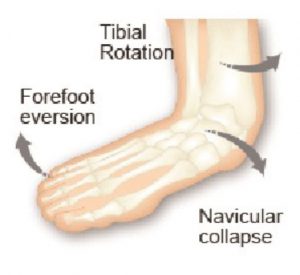
Therefore it can be said that a ‘forefoot varus is a cause of ‘overpronation’ and a forefoot supinatus is the result of ‘overpronation’.1
Merriman’s1 Assessment of the lower limb indicates that:
The Varus foot appears supinated with the lateral border of the foot rela-tively plantarflexed in comparison with the Medial border.
An inverted foot may be due to :
True forefoot varus. Boney abnor-mality, theoretically due to inadequate torsion of the head and neck of the talus during fetal development, but this is not well supported (Kidd 1997)
The presence of a true forefoot varus is said to lead to a very flat foot with no longitudinal arch (Grumbine 1987)
Forefoot supinatus is Acquired soft tissue deformation due to abnormal pronation of the rearfoot. The forefoot Is held in an inverted position be-cause of soft tissue contraction.
It can be difficult to differentiate be-tween a Forefoot Varus and a Fore-foot Supinatus.
The most common test is where a plantar grade pressure is applied to the dorsum of the 1st ray or metatarsal. The 1st Metatarsal shaft should plantarflex and is this does not occur then it is deemed to be fixed osseous condition, whereas if it is mobile or there is some ability to move in a plantarflex direction it a Forefoot Supinatus.
In summary ….a forefoot varus differs from forefoot supinatus in that a fore-foot varus is a congenital osseous deformity that induces subtalar joint pronation, whereas forefoot supinatus is acquired and develops because of subtalar joint pronation.2
Other conditions3 that are due to inverted forefoot are:
A) Dorsiflexed 1st Ray( metatar-sus Primus)
B) Plantarflexed 5th ray both fixed and mobile are possible.3
Assessment :
The Varus foot often looks banana shaped and the navicular has dropped and is excessively everted.
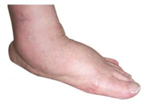
The supinatus foot mimics the Varus foot in most respects, however, the banana shape is not as prevalent and it is able to be distinguished by the ‘Supinatus – Varus test’.
Orthotic Prescription:
Forefoot Varus
When devising the orthotic prescription, firstly the mobility of the rearfoot should be assessed.
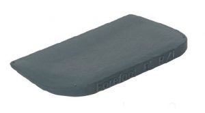
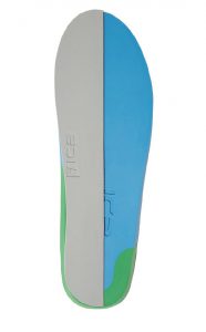
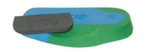
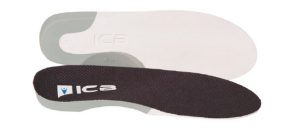
If the Rearfoot is mobile, medium to firm orthotics can be prescribed to support and control the foot, with a forefoot varus addition posted to the medial forefoot.
Alternatively, if the rearfoot is mobile and requires additional inversion (i.e more than the intrinsic posting in the orthotic device) an inversion ramp can be attached to the entire medial aspect of the orthotic.
When the patient has a fixed or arthritic rearfoot, then soft to mid density accommodative orthotics are more effective, with a forefoot varus wedge attached to the medial forefoot. This type of foot can, because of the mobile forefoot, experience conditions such as, Metatarsalgia, morton’s neuroma & mid tarsal periostitis.
Periostitis is an inflammation of the covering of the bones, if left untreated it can progress to a stress fractures.
The Forefoot Varus Addition on the orthotic fills the space under the 1st MTPJ providing the mechanism for toe off to occur by creating normal ground reaction forces to occur at toe off in gait.
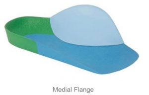
In both cases (i.e. mobile & fixed rearfoot with forefoot varus) extra support can be provided by applying medial flanges (soft or firm) to the dorsal arch area which can also assist in reducing friction on the medial aspect of the foot. Orthotic Prescription: Forefoot Supinatus.
Patient’s exhibiting a forefoot supinatus do not require any additional medial forefoot wedging or modification to the orthotic device .
The reason for this is that once the foot is inside the shoe wear the shoe ‘sock’ upper will assist in a plantarflex action to the dorsum of the foot and this will assist in stretching the contracted soft tissue.
Adding or Posting medial addition or wedge to a SUPINATUS condition will ultimately create a disruption and exostosis between the articulation of the medial cuneiform and base of the first metatarsal at the medial cuneiform joint (1st tarsometatarso joint). .4.
References :
1. Merriman’s Assessment of the lower limb Ed 3 p259-261
2. Forefoot supinatus.Clin Podiatr Med Surg. 2014 Jul;31(3):405-13. doi: 10.1016/j.cpm.2014.03.009. Evans EL1, Catanzariti AR2.
3. Merriman’s Assessment of the lower limb Ed 3 p261
4. Neal’s Disorders of the foot 8th Ed Frowen, O’Donnell,Lorimer,Burrowp126


