Medial Knee Pain

ICB Lower limb biomechanics

Knee pain is a common condition experienced by people of all ages, and levels of activity. Knee pain is a problem that can be experienced due to many contributing factors, Including: increased Q-angle, genu-valgum and genuvarum, muscle tightening through the gastrocnemius, iliotibial band and VMOs, hyper-extension of the knee and tibial rotation and torsion, bakers cyst, connective tissue disorders such as lupus .


View ICB Products

When discussing knee pain, terms such as ‘Retro Patella pain’, ‘Patella Femoral Dysfunction’, ‘Medial Compartment Syndrome’ or ‘Ilio Tibial Band Friction Syndrome’, come to mind. However, from ICB’s experience, these conditions are simply descriptions of knee pain. Knee can be affected by trauma, foot mechanics or issues at the hip. Trauma issues are not discussed in this article , rather we focus on the other two areas, both of which can affect the operation of the knee.

Medial Knee Pain

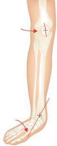
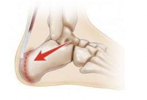
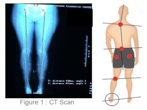
If the patient’s pain is idiopathic (i.e.. no known cause or trauma), excessive pronation and excessive supination may constitute the underlying cause of the problem – as outlined by Michaud, 1997 1. Michaud states that for every 1˚ of pronation the tibia internally rotates 1˚, which in turn impacts on the knee joint, as it takes the stress that is generated by the tibial rotation.

Tibial rotation describes the rotation of the tibial shaft and is different to tibial torsion. Tibial Torsion is a twisting in the osseous structure of the tibial shaft. Internal Tibial rotation is associated with excessive pronation, medially displacing the patello femoral path and encouraging lateral displacement of the patella. Internal Tibial Rotation is a common problem and elicits pain under and around the medial aspect of the patella – especially with excess loading activities such as running and jumping.

Knee Diagram


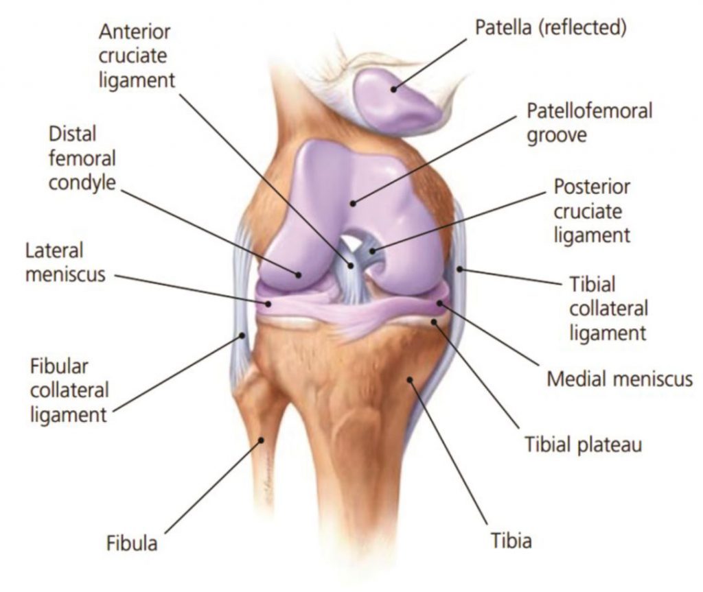
Internal tibial rotation is able to con-tribute to medial collateral strain on the ligament structures that wrap around the medial aspect of the knee and lower leg (Cosgarea, 2002)

To treat pain associated with internal tibial rotation, give VMO strengthening exercises , knee strapping, and other strengthening exercises – all of these are valuable treatment options, however, these methods, no matter how beneficial, only treat the symptoms. Controlling and stabilising the foundation is a key to successful treatment of medial knee pain.

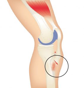
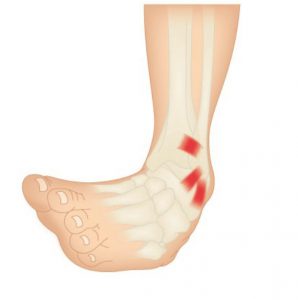
CAUSES OF MEDIAL KNEE PAIN

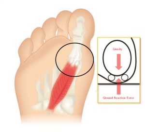
A major cause of medial knee pain is excessive pronation which causes the medial collateral ligament to elongate resulting in the weakening of the VMOs and the ITBs tightening. A con-sequence of this action is external rotation of the femur as compensation. As the ITB and piriformis muscles compensate to reduce medial rotation, the patella begins to track on the lateral aspect of the femoral condyle and a grinding and crackling is felt on the flexion to extension.

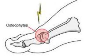
Strapping, using the McConnell technique and strengthening VMOs is a good treatment method, however it will not correct the problem – strapping will only assist in pain relief, and the condition will constantly re-occur until the knee has degenerated or become Osteo-Arthritic, and may eventually require surgical intervention.

A basic requirement to reduce rotational stress on the knee is STJ and MTJ control and correction.

View ICB Products

BIOMECHANICAL TREATMENT

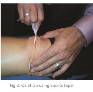
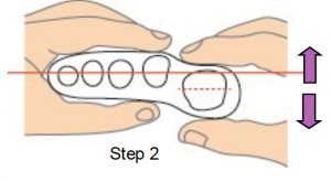
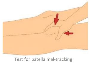
• McConnell strapping technique to control lateral/medial patella displacement, in conjunction with low dye strap-ping to mimic the support and control of an orthotic de-vice. Low dye strapping, however, is only a temporary treatment.

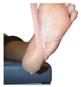
Low Dye taping to imitate wearing orthotics.

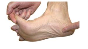
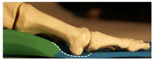
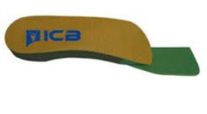
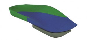
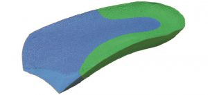
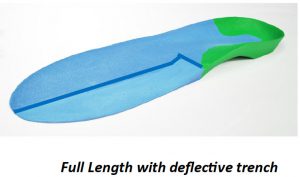
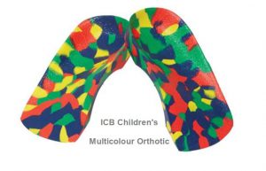
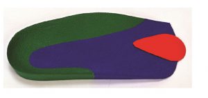
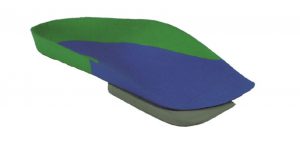
• ICB Orthotics moulded to the patient’s Ideal/NCSP (Neutral Calcaneal Stance Position) to control abnormal STJ and MTJ pronation, by aligning the calcaneus with the lower 1/3 of the tibia and limiting the joint to its original function as a hinge joint.

Calcaneal bisection

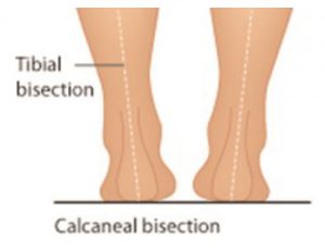
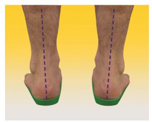
Rearfoot correction using ICB Orthotic with intrinsic 5° Rearfoot varus.

• Strengthening VMO exercises whilst correcting the feet with orthotics, and stretching the ITBs.

• Mobilisation of the knee joint may also be useful.

• Acupuncture can be performed at the point of pain to aid pain relief.

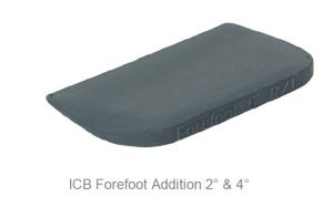
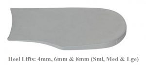
• Runners may require increased rearfoot varus wedgeing to compensate for the higher tibial varum angle at heel strike, during the running cycle.

View ICB Products

REFERENCES:
1 MICHAUD, T.C. (1997) Foot Orthoses and Other Forms of Conservative Foot Care, p10
General References
COSGAREA, A.J., BROWNE, J.A., KIM, T.K. & MCFARLANE, E.G. (2002) Evaluation and Management of the Unstable Patella, The Physician and Sports Medicine, 30, 10
MCCONNELL, J., & COOK, J. (2001) Anterior Knee Pain, Clinical Sports Medicine, 2nd Edition, [http://www.clinicalsportsmedicine.com/ chapters/24a.htm]
PETERSON, L., & RENSTROM, P. (1986) Sports Injuries: Their Prevention and Treatment, Sydney: Methuen Australia

Osgood–Schlatter Disease

ICB Lower limb biomechanics

Osgood–Schlatter disease or syndrome, also known as Apophysitis of the Tibial Tubercle, is an irritation of the patellar ligament at the tibial tuberosity occurring at the tendon-bone junction of the patellar tendon and the tibial tuberosity.1

The condition is named after Robert Bayley Osgood (1873-1956), an American Orthopedic surgeon and Carl B. Schlatter, (1864-1934), a Swiss surgeon who described the condition independently in 1903.

Watch the video below about Osgood–Schlatter disease below.


View ICB Products

General medical opinion is that Osgood Schlatter Disease, is caused by repetitive stress or tension on the growth plate of the upper tibia (the apophysis), which can be complicated by growth spurts and biomechanical deformities or anomalies. In general medical circles it is often called ‘growing pains’ and is similar in bio-mechanical dysfunction to Severs Condition, however occurring in this instant at the tibial tuberosity being characterised by inflammation of the patella tendon and surrounding soft tissues at the point where the tendon attaches to the tibia.

Osgood Schlatter Disease

This condition appears to afflict children who are growing fast and often have external Tibial Torsion condition combined with Internal Tibial Rotation associated with excessive pronation.

Osgood-Schlatter’s Disease, is we believe, caused by repetitive stress or tension on the growth plate of the upper tibia (the apophysis), which can be complicated by growth spurt syndrome and biomechanical deformities or anomalies. The cause is similar in biomechanical operation to Sever’s Disease, except that it occurs at the tibial tuberosity and characterised by inflammation of the patella tendon and surrounding soft tissues at the point where the tendon attaches to the tibia.

As stated, it is usually young people who suffer with this disease – experiencing pain just below the knee joint and patella which usually worsens with activity. It is also associated with an avulsion injury (stretching the tendon so much that it tears away from the tibia and in extreme cases takes a fragment of bone with it – See Figure 2).

Osgood Schlatter’s Condition

A bony bump may appear on the up-per edge of the tibia (below the knee cap) that may be particularly painful when external pressure is applied. It has been misdiagnosed in the past in Australia as “surfer’s knee” (a myth that only surfboard riders suffered from the condition). The hinge motion of the knee is not actually affected.

Most commonly Osgood Schlatters Disease affects active young people, particularly boys between the ages of 10 and 16 who play games or sports that include frequent running and jumping.

In a retrospective study of adolescent athletes actively participating in sports showed a frequency of 21% reporting the syndrome compared with only 4.5% of age-matched non-athletic controls2. Bilateral symptoms are observed in 20–30% of patients indicating that there is a higher incident of unilateral occurrence lead-ing one to deduce that in the unilateral cases, structural leg length or other unilateral biomechanical anomalies may be a contributing factor.

View ICB Products

Symptoms of Osgood Schlatters Disease include:

• Pain over the tibial tubercle
• Swelling over the tibial tubercle
• Weakness in the quad muscle group
• Increased pain & swelling with activity
• A visible lump at the base of the knee cap
• Pain to the touch over the affected area.

Often the pain may last only a few months or may recur until the child stops growing.

Not a lot has been written about the effect of the combination of a growth spurt combined with both pronation or supination, and internal and external tibial torsion. However, children appear to experience more pain and resultant damage to the attachment when these factors occur in combination.

The pain (and resultant effects) become more noticeable during activities that require running, jumping or going up or down stairs and is most common in young athletes who play football, soccer, basketball, netball or who are involved in gymnastics and ballet.

Contributing Factors whilst the Child is still growing

1. Excessive Pronation:
As the foot pronates internal tibial rotation occurs. The body’s mechanism of compensation causes the ITB’s and abductor muscles to tighten in the opposite direction causing the patella to laterally and superiorly displace, hence causing a tractional pull on the tibial tuberosity.

2. Supination & Pronation:

Because of the anterior position of the tibial tuberosity, when the foot supinates due to a FFT valgus deformity less than 10 degrees in the swing phase of gait, the foot lands laterally then the ground reaction forces (on the lateral side) propel the foot into pronation with the same effect above.

3. Internal Tibial Torsion:

Internal tibial torsion will cause the foot to rotate the tibial condyle medially. The ITB’s and gluteals then tighten and come into action as an external rotator of the femur as the compensatory mechanism causing external rotation above the knee and internal rotation below the knee.

The two rotation actions have an effect on the patella and its tendon attachment also causing the tibial tuberosity to stretch and pull.

4. External Tibial Torsion: This causes the foot to externally rotate in the swing phase of gait, the iliopsoas and adductor muscle groups combined to provide compensation at the late swing phase, causing the foot to straighten and land laterally (same effect as 2). Then the ground reaction forces cause the foot to pronate with the help of the psoas pull resulting again in internal rotation above the knee and external tibial torsion position increases tractional forces on the patella and its attachment.

Treatment

• Orthotics moulded to the patient’s Ideal/Neutral Calcaneal Stance Position to realign and hence reduce the effect of tibial rotation.

• Combine orthotic therapy with strapping: use an Osgood Schlatters strap (see below) to reduce the tension on the attachment at the growth plate whilst the child is playing sport (see Figure 3).

Strap Using Sports Tape

Stretching may exacerbate the tearing – alternatively use deep friction massage to help in pain relief.

• R.I.C.E
Active children may experience shortening of the muscles whilst growing, which coupled with biomechanical anomalies = predisposition to Osgood Schlatter.

Contraindications
It is important to differentiate from malignancy, infection, fracture, tendonitis and Sindling-Larsen-Johansson Disease. Initially the diagnosis is based on clinical signs and symptoms including: pain, heat, tenderness and local swelling with prominence at the tibial tuberosity. X-ray is required to establish the extent of the condition.

Steroid injections are discouraged as this may cause weakening of the infra-patellar ligament, scaring and fat necrosis.

View ICB Products

REFERENCES
1 Nowinski RJ, Mehlman CT (1998). “Hyphenated history: Osgood-Schlatter disease”. Am J. Orthop. 27 (8): 584–5. PMID 9732084.
2 Kujala UM, Kvist M, Heinonen O (1985). “Osgood-Schlatter’s disease in adolescent athletes. Retrospective study of incidence and duration”. Am J Sports Med 13 (4): 236–41. doi:10.1177/036354658501300404. PMID 4025675

Hallux Limitus & Rigidus

ICB Lower limb biomechanics

Hallux limitus / rigidus is defined as a degenerative arthrosis of the first metatarsophalangeal joint characterised by a decrease in first metatar-sophalangeal joint range of motion and eventual absence of motion.1


* The Morton’s extension is a 3°- 4˚ ramp under the phalange to allow the patient to propel off at toe off stage of gait. To create a Morton’s extension you can use an ICB 4° Forefoot wedge. and trim.

View ICB Products

The progressive nature of the condition was first documented by Cotterill2 and Davies-Colley3 in literature over a century ago, in which they described hallux limitus / rigidus as a progressive condition producing a severely painful and rigid big-toe joint.

Hallux Limitus is assessed as being evident when movement of the great toe (hallux) is restricted to less than the normal range of motion or flexion to 65 – 75 degrees.


Flexion in the big toe assists to maintain the correct walking gait action to perform normal functions such as stooping down, climbing up, or even standing. For these reasons this disorder can be very troubling and even disabling.

Early signs and symptoms include:
• Pain and stiffness in the big toe
• Abduction of the foot in gait to reduce stress on the 1st MTPJ at toe off.
• Swelling and inflammation around the 1st MTPJ

The main contributing factor biomechanically is as follows:

1. A long 1st metatarsal shaft

2. A short 1st metatarsal shaft In the case of the long metatarsal shaft the 1st MTPJ strikes the ground early and dorsiflexes the 1st metatarsal head. As this happens it restricts the proximal phalange from propelling over the 1st MTPJ and creates an impingement of the joint. Osteoarthritic changes result in the metatarsal joint and narrowing of the joint space leading to hallux limitus or a joint with limited movement. If action is not engaged to prevent it will eventually lead to hallux rigidus or joint ceasure.

A short metatarsal shaft will adduct to the midline of the body to gain ground contact, as this happens the metatarsals will plantarflex and rotate, and the ground reaction forces will cause dorsiflexion of the 1st metatarsal head.

This, when combined with the pronation factor, will create impingement at the tarso metatarso joint and reduce the proximal hallux’s ability to propel over the joint.

View ICB Products
The tarso-metatarsal joint will in this instance be stressed and the ability of the proximal hallux in the propulsive phase of gait to flex to its normal range of motion will be reduced as it impinges or jams the 1st MTPJ.

Often this is referred to as a Functional Hallux Limitus (FHL) as it combines with the pronation factor and ground reaction forces to limit movement. Limited flexion will usually cause 3 types of outcomes:

1.An adductory twist: this is where the calcaneus pivots medially at the propulsive phase of the gait cycle. This is in turn assisted by the external hip rotators such as piriformis, gluteals and ITB’s, and as this takes place medial ground reaction forces (GRF) put pressure on the 1st MTPJ. As the patient attempts to toe off this causes the hallux to deviate and develop me-dial callosity.

2.The distal phalanx of the hallux be-comes the alternative propulsive mechanism which is due to limited joint mobility at the 1st MTPJ, thus causing dorsiflection of the distal hallux with secondary action of nail trauma & nail thickening ( onychogryphosis). The result of this is that many patients wear through the dorsal aspect of their shoe or socks.

3. Abduction of the foot in gait to reduce the impact upon the 1st MTPJ resulting in increased medial pronatory forces.

Correctly aligned and modified orthotic device will alleviate these outcomes and allow the 1st MTPJ to function within the parameters of the structural abnormalities of the foot of the presenting patient.

Once the foot structure is realigned and maintained in the ideal / neutral position pressure is taken off the 1st MTPJ and increased flexion in the joint can be achieved.

Rx : Treatment for a long 1st metatarsal shaft- an orthotic to lift the proximal hallux and at the same time deflect the 1st MTPJ by way of a depression placed under the joint using a heat gun on a 100% EVA orthotic device – using a full length orthotic style is best (see fig. 1 below). Composite orthotic products have limited heat-ing ability and do not allow for easy deflection creation.

Rx: Treatment for a short 1st metatarsal shaft is more complicated as we need to attack the problem on a num-ber of fronts:

1. Correct the pronation factor using an orthotic device moulded to the patient’s ideal / NCSP (Neutral Calacaneal Stance Position) which will control rearfoot eversion (pronation). Generally a device with between 4˚-6˚ rearfoot varus will give the required control.

2. The orthotics should control and correct both the Subtalar and mid tarsal joint pronation together using a device that can be moulded to effectively follow the con-tour of the patients arch to reduce longitudinal arch col-lapse (which would encourage the foot to evert during the propulsive phase) whilst maintaining rearfoot con-trol.

3. Use a ‘Morton’s extension addition’* to lift and sup-port the hallux and allow the hallux to propel over the joint (see Fig. 2). The Morton’s extension lifts the 1st MTPJ to level with the lesser metatarsal joints and allow the proximal phalange to maintain its position and stops the 1st MTPJ from plantarflexing & rotating which encourages the pronation effect.

ICB Green Orthotics

Ultrasound or other physical therapy modalities may be undertaken to provide temporary relief.

Non-steroidal anti-inflammatory drugs (NSAIDs), may be pre-scribed to help reduce pain and inflammation in the 1st MTPJ.

Supplements such as glucosamine-chondroitin sulfate and some vitamins and mineral supplements may also be helpful .

Differential Diagnosis:
Check for gout, rheumatoid and Psoriatic arthritis.
View ICB Products

REFERENCES:
1. MARCINKO DE: Medical and Surgical Therapeutics of the Foot and Ankle, pp 423-465, Williams & Wilkins,Baltimore, 1992.
2. COTTERILL JM: Stiffness of the great toe in adolescents. Br Med J 1: 1158, 1888.
3. DAVIES-COLLEY N: Contraction of the metatarsophalangeal joint of the great toe (hallux flexus). Br Med J 1: 728, 1887.
General references
Camasta C A., 1996 Hallux Limitus and Hallux Rigidus. Clinics in Podiatric Medicine and Surgery. 13(3) pp. 423-445
Chapman C., 1999 Rocker Soles. Podiatry Archives @ www.mailbase.ac.uk/podiatry. January 1999
Chapman C., 1997 Looking through JAPMA. The Journal of Podiatric Medicine. 52(8) pp1113
Durrant M N, Sipert K K., 1993 Role of Soft Tissue Structures as an Etiology of Hallux Limitus. 83(4) pp173-181
Root M L, Orien W P, Weed J H., 1977 Normal and Abnormal Function of the Foot. Clinical Biomechanics Vol 2, Los Angeles
Sanner W H., 1994 Clinical Methods for Predicting the Effectivess of Functional Foot Orthoses. Clinics in Podiatric Medicine and Surgery Vol 2, number 2. pp288-291
Yale, Irving D.P.M ., EdD (Hon.), 1974, Podiatric Medicine

Inversion Sprain and the effects of Forefoot Valgus

ICB Lower limb biomechanics

Orthopedic terminology describes inversion as a frontal plane movement of the foot, where the plantar surface is tilted to face the midline of the body or the medial sagittal plane, the axis of motion lies on the sagittal and transverse planes, a fixed inverted position is referred to as a varus deformity1.

Inversion sprains are probably the most common foot sprain condition and they are often associated with pes cavus feet or a high forefoot valgus in relation to foot mechanics. The treatment of this type of condition will involve a combination of different modalities including orthotic therapy to stabilize the foot mechanics. Watch the video below for a short introduction.


View ICB Products

The most commonly injured site on the foot is the lateral ankle complex, which is composed of the anterior talofibular, calcaneofibular and posterior talofibular ligaments.2,3,4,5

This article will focus on lateral sprain and pain associated with a pes cavus foot structure and a forefoot valgus deformity.

Inversion Sprain

Repetitive lateral ankle sprain or lateral knee pain (or even lateral shin splints) is often diagnosed as ‘idiopathic’, closer examination of the biomechanical relevance needs to be pursued. The term ‘idiopathic’ is often used in this area as there seems no reason for the pain occurrence. The meaning of idiopathic is a derivative of the Greek word idios “one’s own” and pathos “suffering”, or ‘arising spontaneously or from an obscure or unknown cause’ 6


Pes cavus foot (high arch) structures (see Figure 1) appear to have a predisposition to lateral ankle sprains and present as a rigid structure and a supinated foot structure.
Pes Cavus foot Structure

Patients with this foot structure will often complain that their joints are painful and when they walk without shoes on hard floors.

Anterior view Supinated foot using ICB AAM line

This type of structure will usually exhibit a forefoot valgus deformity meaning that:
‘The plantar plane of the forefoot remains everted relative to the plantar plane of the rearfoot when the subtalar joint is in neutral.’

This deformity will have an impact on the patient in heel strike, midstance and toe off phase of gait. The patient who exhibits a pes cavus foot structure will often present with a (FFVL) Forefoot valgus greater than 10º and will often exhibit a plantar flexed 1st metatarsal (Boyd & Bogdan, 1993), encouraging the foot to strike laterally and eliciting pressure on the lateral aspect of the hip joint.

If the Forefoot valgus deformity is greater than 10º, the foot will often continue to supinate through the cycle, having a jar-ring effect on the upper structure, putting additional strain on the Lateral aspect.
Biomechanical protractor

View ICB Products

When the foot is supinated it will stress on the peroneals and may cause elongation of the muscles and tendons, thus weakening the retinaculum and lengthening the peroneals and may cause the tendon to sublux off the lateral aspect of the malleolar.

The FFT Valgus deformity (in gait) encourages the foot to invert the foot, propulsion is delayed causing lateral instability and results in tension and tearing of the peroneal muscles, causing inflammation and tenderness, and difficulty walking. Lateral ankle sprains are more common than medial due to the fact that ligaments are weaker on the lateral side7.

Hence the lack of lateral stability can be caused by uncompensated or partially compensated rearfoot, a flexible forefoot valgus or osseous forefoot valgus (Boyd & Bogdan, 1993; Hollis et al, 1995; Shapiro et al 1994).

There are also certain biomechanical foot deformities that make some patients more susceptible to inversion sprains, such as, neurological deficits and supinated foot types which exhibit or function with a supinated calcaneus (Valmassy, 1996),

In gait the FFTVL allows inversion of the foot to gain ground contact.

Inversion Sprain

ICB Orthotics

In summary, if a patient presents with lateral hip pain, knee pain, ankle strain or repetitive lateral inversion sprain, check for a forefoot valgus deformity and employ the following treatment suggestions to assist in alleviating the pain symptoms, whilst treating the underlying causation.

If the patient requires a forefoot valgus posting to be added to their orthotic devices remember that the measured forefoot valgus deformity commence by taking ½ the measured amount and gradually increasing until treatment is effected or the patients shoe wear is not able to accommodate.

ICB Forefoot Addition

If the forefoot valgus is very large say greater than 15º, a reduction in the posting size may be required to assist the patient in being able to accommodate the orthotic in the shoe box area.

Suggested Treatment Options for Lateral Pain:

• Deep tissue massage along the peroneal muscles or suction cups to break down scaring and adhesion.

• Orthotic device with an appropriately sized forefoot valgus addition applied (see Figure 4). Always start with a conservative sized valgus addition, and build up from there to assist compliance.

• Acupuncture at the point of pain.

• Lateral Prolotherapy to strengthen the lateral ankle ligaments to encourage proliferation of the lateral ligaments (formation of collagen fibres).

• Mobilisation of the cuboid joint, as this may become subluxed.

• For lateral ankle sprains use stabilising ankle strapping, in combination with the orthotic device.

The USA National Athletic Trainers’ Association (NATA) new guidelines for treating and preventing ankle sprains in athletes are as follows:

• Early use of non-steroidal anti-inflammatory drugs (NSAIDS) post injury
• Functional rehabilitation rather than immobilisation for grade I and II ankle sprains
• Prophylactic ankle supports for athletes with a history of previous ankle sprains
• Immobilisation with a rigid stirrup brace or below-knee cast is recommended for grade III sprains for at least 10 day followed by controlled therapeutic exercise.
View ICB Products

REFERENCES :

1.McGraw-Hill Concise Dictionary of Modern Medicine. © 2002 retrieved 11.09.14
2.Ivins D. Acute ankle sprain: an update. Am Fam Physician. Nov 15 2006;74(10):1714-20. [Medline].
3.ANDERSON KJ, LECOCQ JF, CLAYTON ML. Athletic injury to the fibular collateral liga-ment Of the ankle. Clin Orthop. 1962;23:146-61. [Medline].
4.Gross MT, Liu HY. The role of ankle bracing for prevention of ankle sprain injuries. J OrthopSports Phys Ther. Oct 2003;33(10):572-7. [Medline].
5.LeBlanc KE. Ankle problems masquerading as sprains. Prim Care. Dec 2004;31(4):1055-67. [Medline].
6.Merriman and Webster Medical Dictionary retrieved 2/10/14
7.M. A. R. Freeman Lateral Ligament of Ankle JBJS Vol. 47 B, NO. 4, Nov 1965
8.Kaminski TW, Hertel J, Amendola N, et al. National Athletic Trainers’ Association position statement: conservative management and prevention of ankle sprains in athletes. J Athl Train. 2013;48(4):528-45.
General references
BOYD, P.M. & BOGDAN, R.J. 1993. Sports Injuries. In LORIMER, D.L. Neales Common Foot Disorders: Diagnosis & Management: A General Clinical Guide (4th Ed). Churchill Livingstone, Edinburgh, P. 179-180
HOLLIS, J.M., BLaSIER, R.D., CHARELENE, M.F. & HOFMANN, O.E. 1995. Biomechanical Comparison of Reconstruction Techniques in stimulated lateral Ankle Ligament Injury. The American Journal of Sports Medicine, 23, (6), p.678-682
SHAPIRO, M.S., KABO, J.M., MITCHELL, P.W. & LOREN, G. 1994. Ankle Sprain Prophylax-is: An Analysis of the Stabilising Effect of Braces and Tape. The American Journal of Sports Medicine, 22, (1), p 78-82.

Sesamoiditis

ICB Lower limb biomechanics

Sesamoiditis is painful inflammation of the sesamoid apparatus, which is located in the forefoot under the 1st MTPJ. It is not a particularly common condition and can be at times difficult to diagnose and treat. Generally it is accepted that the sesamoids perform two principal functions:

1. Absorbing impact forces in the fore-foot during walking through a series of attachments to other structures in the forefoot.

2. Acting as a fulcrum to provide the flexor tendons with a mechanical advantage as they pull the hallux down against the ground during gait.

Watch the short video below

The hallux (big toe) has two phalanges and two joints (interphalangeal joints), together with two small sesamoid bones (the medial and lateral sesamoid bones).

The sesamoids are implanted or embedded in the flexor hallucis brevis tendon which exerts pressure from the big toe against the ground and aids in the act of walking during the toe-off phase of gait. The sesamoids not only have to endure the pressure of body, weight and gravity, but also the constrictive pressure of the flexor hallucis brevis tendon.

There are generally five categories of sesamoid apparatus type injuries:

A. Sesamoiditis is a general description of the condition which is given to inflammation of the sesamoids but actually relates more specifically to tendonitis of the flexor halluces longus1.

B.  Stress fractures of the sesamoid usually occurring as an impact injury.

C. Acute sesamoid fracture most commonly an evulsion fracture.

D. Osteochondritic dissecans or avascular necrosis. A feature in which the blood supply to the sesamoid bone is interrupted in which bone death can result.

E. Chondromalacia of the sesamoid resulting from continual synovitis of the 1st MTPJ in which fibrosis develops leading to freezing of the sesamoids1 and apparatus.

View ICB Products

Sesamoiditis is the most common and is characterised by pain at the first metatarsal sesamoid commonly occurring in combination with a plantar flexed 1st metatarsal. This condition may present as often as in 4% of overuse type foot injuries (Dennis and McKinney, 1990).

Sesamoiditis typically affects physically active young people and is more common in sporting people who require to balance on the ball of the foot (such as ballet dancers, basket ballers, netballers, cricket players and soccer players) – as they attempt to stop and change direction by pivoting or putting pressure on the forefoot area and the 1st MTPJ.

Sesamoiditis Pain

The condition causes pain under the 1st MTPJ and commonly affects the medial (inner) side of the joint. The pain may be constant, or it may occur with or be aggravated by, movement of the big toe joint. It may also be accompanied by swelling (oedema) throughout the plantar aspect (bottom) of the forefoot.

It is reputed that the forefoot bears approximately one half the body’s weight and balances pressure on the ball of the foot. Sesamoiditis has been also linked as a possible uncommon cause to Metatarsalgia2.

It is generally accepted that the sesamoids perform two principal functions:

1. Absorbing impact forces in the forefoot during walking through a series of attachments to other structures in the forefoot.

2. Acting as a fulcrum to provide the flexor tendons with a mechanical advantage as they pull the hallux down against the ground during gait.

Patients who suffer from Sesamoiditis often exhibit a fixed or mobile plantar flexed 1st metatarsal which maintains the 1st MTPJ on impact. The lesser metatarsals absorb the impact in the gait cycle, however, if the 1st MTPJ is rigid and the sesamoid take the full impact during toe off. Repetitive chronic pressure and tension on the forefoot will cause the surrounding tissues to become irritated and inflamed. In some cases the sesamoids may bifurcate and in severe cases necrosis may occur.

The practitioners should check the patient for a Plantar flexed 1st using the common test in which the movement of the joint should be 5mm dorsiflexion, and 5mm plantar flexion or a total range of approx 10mm.

This test will identify if there is a limitation in the joint’s range of motion due to a fixed osseous condition.

How to perform this test:

1.With the patient in the supine position, maintain the foot in the neutral position, using ICB Anterior Alignment Method.

2. Grip the lesser metatarsals, line the thumbs up and the range of movement should be 5mm up and 5mm down.
Step 2

Pronation will displace the sesamoids laterally causing trauma to the sesamoid apparatus. This can change the patient’s gait as they try to compensate and may cause other upper body compensatory effects, including hip pain.

A Supinated foot will often display a plantar flexed 1st as compensation to assist in lowering the 1st MTPJ to the ground and thereby enabling toe off.

Patients who suffer from Sesamoiditis may exhibit a fixed or mobile plantar flexed 1st metatarsal 3 which maintains the 1st MTPJ in a plantar flexed position on impact. The lesser metatarsals are able to absorb the impact in the gait cycle, however, the 1st MTPJ is rigid and the sesamoid take the full impact during toe off.

Repetitive chronic pressure and tension on the forefoot will cause the surrounding tissues to become irritated and inflamed. In some cases the sesamoids may bifurcate and in severe cases necrosis may occur.

Treatment

Initial treatment for sesamoiditis is usually non-invasive and includes orthotic therapy to treat the plantar flexed 1st. Generally a deflection will need to be incorporated into the orthotic to remove or reduce the pressure from this area.

ICB Orthotics

The Plantar flexed 1st Ray (metatarsal) deflection is cut around the 1st MTPJ and then the edges are heated and smoothed to taper the edge of the orthotic, and avoid irritation (see opposite). The orthotic itself has some in-built forefoot support under the 2nd to 5th to support this area similar to a 2-5 bar.

Treatment is generally non-surgical, however, if conservative measures fail, surgical intervention by way of removal or pinning of the sesamoids may be undertaken.

Modifying the patients’ shoes is not overly effective as each pair would need to be modified. The easier approach a heat mouldable orthotic incorporating a Plantar flexed 1st ray deflection modification which can be moved and transferred from shoe to shoe with relative ease.

In addition, the big toe may be bound with strapping tape (or athletic strapping) to immobilise the joint as much as possible and allow healing to take place.

Acupuncture can be used to reduce inflammation and anti inflammatory drugs can be taken to reduce swelling.

If a full length orthotic is required a ‘relief trench’ can be placed into the orthotic using a dremel hand grinder to allow the 1st MTPJ to plantar flex without restriction.

Full Length Orthotic with deflective trench

Differential Diagnosis:

• Stress fracture of the sesamoid bone

• Avulsion fracture or sprain of the proximal pole of the sesamoid

• Sprain of the distal pole

• Sprain of a bipartite sesamoid

• Arthrosis of the sesamoid metatarsal articulation. Arthrosis pain is often felt during the day and emphasises during joint movement efforts

• Pre-radiographic osteonecrosis of the sesamoids

View ICB Products

References
1 Neal’s Disorders of the foot 8th edition 2002 p318
2. Merriman’s Assessment of the Lower Limb 3rd Edition. Churchill Livingstone. p331
General REFERENCES
Michaud T.C. (1993) Foot Orthoses and Other Forms of Conservative Foot Care. Williams and Wilkins, Baltimore, pp.93-105.
Root M.L., Orien W.P. and Weed J.H., (1971) Clinical Biomechanics: Normal and Abnormal Function of the Foot. Clinical Biomechanics Corp, Los Angeles, Chapter 10.

Severs Condition

ICB Lower limb biomechanics

Severs Condition (Calcaneal Apohphysitis) is an inflammation of the growth plate of the bone at the back of the heel (apophysitis of the calcaneus) and occurs during a child’s adolescent years, usually affecting children who are physically active. The point where the inflammation of Severs condition appears is usually localised to the posterior and plantar side of the heel over the calcaneal apophysis – where the Achilles tendon attaches to the back of the calcaneus.
Watch the short video below:


View ICB Products

Often young children experience pain in one or both heels whilst engaging in general activities such as running, jumping, or sports such as netball, basketball, football and soccer. Symptoms of Severs Condition include extreme pain when the child places their heel on the ground. This pain is alleviated when the child runs or walks on their toes (i.e. activities that do not exert weight and pressure on the heels). This is because strain is reduced on the epiphyseal plate as the heel is raised off the ground.

The calcaneus takes on a slightly supinated position thus reducing tension on the attachment.

Often full immobilisation is recommended to treat this condition, however, there is no need to plaster the foot or for the child to stop playing sport during the course treatment. That is, unless X-rays show evidence of a fracture in the heel growth plate or epiphyseal plate. (see Fig:1) If a fracture is evident, sporting activities should cease for approximately 6 weeks to allow healing.
X-ray Severs Condition

This condition usually affects children, between the ages of 8 to 13 years. Severs condition is more common in boys than in girls, and it is uncertain why this appears to be the case. Micheli and Ireland reported on 85 patients, 64% of whom were male1 in their research – however, they did not provide definitive reasons for the skew towards young males.

In one prospective study of injuries among players aged 9-19 years in football academies, 2% of overall football injuries were due to Sever’s Condition; the peak for incidence was in the under-11 age group2. In a study of 85 children, the condition was bilateral in 61%.3

General research opinion agrees that between the ages of 8 and 13 years, children’s bones are still in the growth stage and the growth plates on the skeletal structure have not yet become fused and ossified, which normally occurs by around the age of 17 years4. During the rapid growth surrounding puberty, the apophyseal line appears to be weakened further because of increased fragility of the calcified cartilage.

The combination of growth and poor foot function and mechanics, can cause inflammation and pain at the point where the
tendo-Achilles inserts onto the calcaneus.
Severs Condition

Excessive pronation is a major contributing factor when combined together with the child’s growth spurt pattern.

In very severe cases, the growth plate can detach from the calcaneus. Fractures or micro fractures can completely detach the growth plate, pulling it out of position possibly resulting in osteochondritis dissecans.

Neal’s Disorders of the Foot states that, “Repetitive contractions of the muscles of the posterior compartment (gastrocnemius and soleus) may predispose to micro-fractures at the calcaneal epiphyseal plate5.”

Severs Condition Squeeze test

Assessment Techniques

Sever’s condition can be diagnosed based on the child’s medical history and presenting symptoms. A simple and effective test is the calcaneal “squeeze test” which will identify localised pain symptoms and then this will need to be combined with other tests to confirm the diagnosis. Once the squeeze test has been performed practitioners will need to assess for:

* Decreased ankle dorsiflexion.* Abnormal hind foot motion (eg.excessive pronation or supination).* Tight calf muscles* Excessive weight-bearing activities e.g running which will cause deformation of the rearfoot.

Madden and Mellion (1996) noted an means of assessment could be to require the patient to stand on their tip toes, raising and lowering, and gauge whether there was an exacerbation of symptoms6.
Childrens Orthotics
View kids Orthotics

View ICB Products

Treatment

Orthotic therapy is an excellent treatment regime7 as it realigns the biomechanical structure whilst controlling excessive pronation of the rearfoot.

Stabilising the calcaneus and its growth plate by restricting or limiting excessive pronation can prevent long-term injury and discomfort.

The use of bilateral heel lift of approximately 4mm is recommended in combination with orthotics for short term relief. Removal of the heel lifts, after an initial 2 week acute pain period is recommended, however if additional support and control is required sports strapping using a low dye taping technique can be useful.

ICB Heel Lifts

Deep tissue massage of the calf muscle (in combination with orthotic therapy), is also a successful mode of treatment. Stretching the calf muscles is however not recommended whilst inflammation is still present, as this may cause further tearing at the attachment. Stretching and deep tissue massage may be in part, prophylactic in the maintenance of the condition.

View ICB Products

REFERENCES
1 Leri JP; Heel Pain in a Young Adolescent Baseball Player. (2004) Journal Chiropractic Medicine. Spring: 3(2):66-8.
2 Price RJ, Hawkins RD, Hulse MA, et al; The Football Association Medical Research Programme: An Audit of Injuries in Academy Youth Football. Br J Sports Med. 2004 Aug;38(4):466-71.
3 Micheli LJ, Ireland ML; Prevention and Management of Calcaneal Apophysitis in Children: An Overuse Syndrome. Journal Paediatric Orthopaedics. 1987 Jan-Feb;7(1):34-8.
4 Noffsinger MA, (Feb 2012) Sever Disease, Medscape
5 Neal’s Disorders of the Foot 8th Edition p113
6 Neal’s Disorders of the Foot 8th Edition p114
7 The American College of Foot & Ankle Orthopedics & Medicine, Prescription Custom Foot Orthoses – Practice Guidelines, December 2006

Back Pain and Leg Length

ICB Lower limb biomechanics

Back pain is a complex one as it can be categorised into a variety of locations Upper, Middle, and Low Back Pain, and can range in intensity from mild to severe (chronic). Sciatica for instance, can cause pain in the lower back and the legs, where-as low back strain can affect L1 –L5 region of the spine. Excessive pronation can also cause lower lumbar pain due to its affect of rotating the pelvis anteriorly. A short video on Back pain is below.

View ICB Products

Treating this pain requires that we determine wherever possible, the origin of the disorder whether from the muscles, nerves, bones, joints or other structures in the spine.

It is reported that low back pain causes 40% of missed days of work in the United States1, and is reputed to be the single leading cause of disability worldwide2.

The spine is a complex interconnecting network of nerves, joints, muscles, tendons and ligaments, all of which are capable of producing pain. Therefore in this newsletter we just want to focus on basic back pain associated with structural leg length abnormality and how to treat these problems by incorporating orthotics and other modalities.

Leg length can, we believe, be categorised into two basic areas:

 1. Structural – a difference in the long bone measurement of the leg and,

 2. Functional – arising from a variety of causes which affect the biomechanics of the body.

When a patient presents with back pain a determination of the underlying factors needs to be undertaken. Always start with the basics, and complete a full biomechanical assessment, which includes long bone measurement and a comparison of the amount of pronation/eversion and supination/ inversion at the foundation. The first thing to look for is whether the patient is presenting with bilateral or unilateral pronation. If it is UNILATERAL, establish if the patient has:

a. suffered a trauma in which one plantar fascia may have ruptured or elongated and consequently, created a functional short leg or if there is idiopathic ulilateral excessive pronation or supination present.

b. assess if tibial torsion with hip compensation is present : such as tight psoas which will rotate the pelvis anteriorly, or tight piriformis ITB’s and Gluteals, as a compensation for internal tibial torsion to correct the pigeon toed position. Both these compensations could either, rotate the pelvis (anterior) forward or (posterior) backward creating functional leg length problems in the gait cycle.

Cat Scan

If the patient has a long leg compensation in which the long leg drops down or pronates to level the pelvis, establish the structural length and this can be verified using a CT scanogram (see Figure 1) for leg length or the normal x-ray film to check the leg length.

The longer leg will be predisposed to hip joint wear and tear. Therefore if a patient is asymmetrical, inform them about the consequences of not treating the condition. Some practitioners say the body will compensate for the difference so why bother with a heel lift to raise and level the pelvis? However, even everyday objects that are asymmetrical such as tables and chairs become unstable over time, how much more our bodies?

It has been said that 3-4mm can upset the biomechanical structures of the body.

If the patient is suffering from hip compensations they will need to be either referred to the appropriate Allied Health Practitioner for stretching and strengthening exercises to correct the anomaly. At the same time prescribe orthotics to realign and correct the biomechanics. A combination of orthotic therapy and muscle stretching and strengthening will give the best treatment results.

If there is no apparent structural leg length the use of orthotics to realign the foundation will be required and a combination of other modalities will be needed to address both skeletal and soft tissue imbalances and compensations.

When the patient presents with a structural deformity either due to a recent trauma or a long term biomechanical related anomaly, the use of a heel lift or raise on the structural short leg will be required.
ICB Heel Lift for ICB Orthotics

It is recommend that the practitioner applies ½ the leg length discrepancy measurement, in the heel lift. Then this amount can be gradually increased, this will limit initial discomfort and quicken patient compliance. In the case of treating a short leg due to trauma/ accident injury the entire amount can be applied, within the first 6 months, as long as the rehabilitation has ceased and the body has not started to use soft tissues to compensate for the leg length deficiency. Contraindication in these instances would be any fusing of the spine as this may cause the patient to be in more pain.

If you are prescribing orthotics and have NOT MEASURED structural leg length, the orthotic will invariably remove the any normal body compensation of the ‘long leg pronation’. If excessive long leg excessive pronation compensation is removed it will create a jamming in the long leg acetabulum at the hip, create problems with the SI joint and contribute to the development of a scoliosis, as stated in Blake & Ferguson, 1992.

Placing a heel lift in the shoe of the short leg without using orthotic devices is not recommended, as this will create a leg length problem on the raised leg side, because the longer structural leg will continue to pronate due to ligament laxity. The use of one orthotic is never recommended as the foundation needs to be balanced, and the necessary biomechanical corrective additions attached to the orthotic.

Where back pain and SI joint pain is concerned it is recommend that leg length discrepancy is assessed and treated.

View ICB Products


Treatment
Measure and confirm structural or functional leg length discrepancy. Use a method you are comfortable with.
Medial Malleoli symmetry

• Orthotics to treat ‘long leg pronation’, together with a heel raise being added for the short leg.

• Raise the short leg in stages – if the discrepancy is a large, start with half the amount of measured discrepancy.

• Often a multi-disciplinary approach such as adjustments to ‘free’ the pelvis, stretching tight muscles is beneficial. Check the head of femur is sitting correctly in the acetabulum, in combination with orthotic therapy will deliver the best results for the patient.

View ICB Products

REFERENCES:

1. Manchikanti, L; Singh, V; Datta, S; Cohen, SP; Hirsch, JA; American Socie-ty of Interventional Pain, Physicians (Jul–Aug 2009). “Comprehensive review of epidemiology, scope, and impact of spinal pain.”. Pain physician 12 (4): E35–70. PMID 19668291.
2. Institute for Health Metrics and Evaluation. “2010 Global Burden of Disease Study”
3. Gary Fryer 2005 : Factors affecting the intra-examiner and inter-examiner reliability of palpation for supine medial malleoli asymmetry.
General REFERENCES:
Blake, R.L. and Ferguson H (1992) ‘Limb Length Discrepancies’ Merriman, LM and Tollafield, DR (1995) ‘Assessment of the Lower Limb’
Blake RL, Ferguson, H (1992) Limb Length Discrepancies. Journal of the American Podiatric Medical Association 1: 33-38.
Blustein SM, D’Amico JC (1985) Limb Length Discrepancy –identification,Clinical Significance and Management. Journal of the American Podiatric Medical Association 75(4): 200-206.

Shin Splits

ICB Lower limb biomechanics

Shin splints or Tibial Fasciitis1 (TF) is a descriptive term for pain in the soft tissue apparatus of the tibia, medial pain being the most common and is referred to by American Academy of Orthopaedic Surgeons as Medial tibial stress syndrome (MTSS) being described by the academy as “pain along the inner edge of the shin-bone (tibia).”

Shin splints are a common injury affecting athletes and others who engage in running sports or other forms of physical activity. Characterised by general pain in the lower region of the leg: between knee and ankle.

Recent reports suggest that it comprises 13% to 17% of all running-related injuries 2,3 .

Often patients experience shin splints after being told by their General Practitioner to start walking as a means of healthy cardiovascular exercise.

Shin splints are classified as an over-use syndrome due to fatigue failure at the deep fascial attachment sites of the tibia, excessive supination or excessive pronation is often an underlying causative factor. Excessive supination and the association of forefoot valgus has been linked to the lateral shin splints, whereas excessive pronation is more often at tributed and associated with medial shin splints.

At mid stance, with the foot excessively pronated and the tibia internally rotated, the tibialis posterior muscle and tendon is placed under tension at its attachment to the tibia and the interosseous membrane. Repetitive stress initially produces tendonitis. The tibialis anterior and tibialis posterior muscle is partly responsible for inverting the foot and ankle in respect to the midline of the body and assisting in plantarflexion of the foot. Another function is that it helps to maintain the arch of the foot and is particularly active during weight-bearing activity such as walking and running.

When training it is NOT advised that one should train through the ‘pain barrier’ of the initial shin splint type injury, as the chronic traction may produce periostitis. (Bruhner & Khan, 1993; Lorimer et al, 1997).

Pain in the shin can be separated into 3 areas:

1. Medial shin pain
2. Lateral shin pain
3. Anterior shin pain

Medial Shin Pain: usually occurs when the patient engages in prolonged walking or running uphill. The patient will feel the pain along the tibialis posterior muscle and tendon. Sometimes it can be experienced close to the inferior medial collateral ligament attachment, all the way down to the navicular area.

The Tibialis Posterior tendon may slip over the medial malleolar due to tight Tibialis Posterior tendon, this action can be attributable to a weak retinaculum. Repetitive STJ pronation, causes the Tibialis Posterior tendon to elongate as the foot collapses medially, hence causing medial shin pain. Constant elongation may contribute to the development a tears and scaring in the muscle belly, and a pulling away from the tibia, causing inflammation and pain.

Differential Diagnosis: Compartment Syndrome and stress of the tibia.

Treatment for Medial Shin Pain

• R.I.C.E technique
• Deep tissue massage therapy – to break down adhesions.
• Orthotic therapy to control the excessive pronation.
• Low dye strapping to mimic orthotic and give additional support (short-term treatment only).

Lateral Shin Pain: the patient may feel this pain when undertaking excessive running and walking activities and is mainly experienced when walking or running downhill. The biomechanical aspects of the foot may be a high forefoot valgus or fixed plantarflexed 1st ray, both of which may cause a supinatory effect.

In gait, contraction of the anterior tibialis muscle raises the toes and dorsiflexes the foot. The main functions of the anterior tibialis during athletic activities are dorsiflexion through concentric contraction, and slowing the reverse movement, plantarflexion, through eccentric contraction.

Weakness of the anterior tibialis, particularly in eccentric contractions (lengthening contraction), can cause cumulative damage to the muscle and surrounding connective tissue.

Strength of the posterior muscles of the lower leg is an-other factor implicated in both anterior shin splints and posterior shin splints. Though it is less common in the case of anterior shin splints, a lack of support from the calf muscles may contribute to shin splint pain. (see Fig. 1).

In addition to this, if the foot exhibits a forefoot valgus, the foot in gait seeks to reach the ground and it does this by encouraging the foot into supination, hence tearing the peroneal muscles and causing inflammation and tenderness to touch, and difficulty walking. See Figure : 2

Differential Diagnosis: Compartment Syndrome and stress of the tibia.

Treatment for Lateral Shin Pain

• R.I.C.E.
• Strapping with a valgus wedge added for short term relief.
• Orthotic therapy (and the addition of lateral wedging ) to control the supination.

• Add a 1st ray cut-away to the orthotic if the patient has a plantarflexed first ray (see Figure 3).

Anterior Shin Pain: the patient normally feels the pain after running or walking and is mainly associated with uphill and downhill walking and running.

Biomechanically, it is related to supination at heel strike and then pronation at the mid stance to toe off stage.

During the gait cycle the Tibialis Anterior muscle acts as an inverter of the foot to allow for ground clearance at late swing phase. If there is a forefoot valgus of <10˚ (less than) the foot will supinate at heel strike and the ground reaction force on the lateral side will then propel the foot into pronation at mid stance of gait to toe off. In this circumstance the Tibialis Anterior will elongate and cause tearing to occur at the muscle belly especially on the lateral tibial crest and cause adhesions.

In short, during swing phase the tibialis anterior contracts causing the foot to invert and dorsiflex. At lateral foot strike the ground reaction forces propel the foot into pronation in early midstance to late midstance, causing a sudden pull of the tibialis anterior. This firstly causes a shortening of the tibialis anterior, then a sudden lengthen-ing of the tibialis anterior from midstance to the toe-off stage.

Differential Diagnosis: Compartment Syndrome and stress of the tibia.

Treatment for Anterior Shin Pain

• R.I.C.E. technique.
• Deep tissue massage at the Tibialis Anterior and along the lateral tibial crest.
• Suction cup therapy
• Acupuncture on the adhesion to help break down scaring.
• Orthotic therapy – not only to control pronation, but also the supination at heel strike and the a forefoot lateral wedge to treat the Forefoot Valgus if apparent.

References
1 Steven I Subutnick. (1999) Sports Medicine of the Lower Extremity p277
2 Clement D., Taunton J., Smart G. (1981). “A survey of overuse running injuries”. The Physi-cian and Sports Medicine 9, 47–58.
3 Cox J. S., Lenz H. W. (1984). Women midshipmen in sports. American Journal of Sports Medicine, 12(3), 241 – 243.
General References:
BRUNKER, P., & KHAN, K. (1993) Clinical Sports Medicine, Sydney: McGraw-Hill Book Com-pany
LORIMER, D., FRENCH, GWEN, & WEST, S. (1997) Neales Common Foot Disorders: Diagno-sis and Management, 5th Edition, Melbourne: Churchill Livingstone

Metatarsalgia: Ball of Foot Pain

ICB Lower limb biomechanics

Metatarsalgia is a general term used to denote a painful foot condition in the metatarsal region of the foot, the area just before the toes, more commonly referred to as the ball of the foot. Thomas G. Morton ( I867) is credited with identifying the condition1. This is a common foot disorder that can affect the bones and joints at the ball of the foot. Metatarsalgia (ball of foot pain) is often located under the 2nd, 3rd and 4th metatarsal heads, or more isolated at the first metatarsal head (near the big toe). Differential diagnosis of Metatarsalgia is Morton’s Neuroma which exhibits localised pain than Metatarsalgia.


This as often it affects the sufferer’s quality of life. Metatarsalgia restricts the distances patients can walk, and causes extreme pain and discomfort. It is a simple condition to treat effectively, however left untreated, this condition can be debilitating to its sufferers.

Typically the patient will present with an ‘all over burning or stone bruise feeling’, often the foot will feel swollen.

There are many contributing causes for the condition, the main one being excessive pronation. Other issues such as diabetes, Rheumatoid Arthritis, gout and excess fluid build up can all provide the environment to promote the development of increased stress on the ball of the foot resulting in Metatarsalgia.

Most articles written about the condition appear to combine Metatarsalgia and Morton’s Neuroma, whereas there are two distinct conditions, one a generalised all over pain or feeling of discomfort and the other (Morton’s neuroma) a specific localised pain usually presenting between either the 2nd and 3rd or 3rd and 4th meta-tarsal joints.

The more common and certainly the easier condition to address is Metatarsalgia in which excessive pronation appears as the main underlying causative factor. General podiatric opinion appears to credit the cause as ‘a forced change of the dynamics of the foot’ as outlined by ACFAOM (American college of Foot Orthopedics & Medicine). Increased obesity is often a ‘trigger’ as it places additional stress on the foot structure.

Excessive pronation for any length of time can cause weakening of the soft tissue structures and as a result ligamentous laxity and muscle wastage will occur. As this occurs in the forefoot, the metatarsals plantar flex and rotation takes places resulting in the loss of the transverse arch.

These shearing forces on the forefoot structures causing pressure and pain.

Long term damage to the nerves may be caused and callosities will be exhibited in the patient. With this condition one or more of the metatarsal heads become painful and/or inflamed, usually due to excessive pressure over a long period of time. It is common to experience acute, recurrent, or chronic pain with Metatarsalgia. Also, as we get older, the fat pad in our foot tends to thin, making us much more susceptible to pain in the ball of the foot (Cailliet, 1983; Lorimer et al, 1997).

Treatment

Research has indicated that nonsurgical management is usually sufficient to achieve satisfactory results2 when dealing with Metatarsalgia. We should be encouraged to pursue conservative treatments as a first approach after establishing the underlying causative factors such as mechanics, foot anatomy, and foot and ankle deformity.

1. Identify if the feet are pronating or supinating.

2. If pronation exists then place the foot into the ideal or subtalar neutral position, and identify the amount of pronation, i.e. slight, moderate or severe. This will help you identify the appropriate ICB Orthotic type to heat mould – then mould the orthotic to the subtalar neutral joint position(STJ). It is considered that mild pronation needs less support, whilst severe pronation requires firmer orthotic support.

3. Don’t forget to consider the patient’s body weight also – light weight patients need less control than overweight patients.

If the patient has been excessively pronating for a long time, not only will they have lost the longitudinal arch but they may have also lost the transverse arch.

This situation will require that the practitioner heat moulds to the patient’s neutral calcaneal stance position, and will then apply a metatarsal dome to restore the transverse arch.

The size of the dome will depend on the width of the foot – i.e. a narrow foot will only require a small metatarsal dome, a wide foot, a larger dome. The function of the metatarsal dome is to promote the transverse arch and separate the metatarsal heads to prevent nerve impingement and entrapment.

If the foot is supinating or the patient has a high Pes Cavus foot type this allows increased pressure on the metatarsal heads and callous formation. Always check for a forefoot valgus deformity greater than 10˚. A plantarflexed 1st ray (metatarsal) can also elicit a lateral shift as compensational repositioning of the foot and may present with a forefoot valgus anomaly together with a pes cavus foot type and often clawed toes will be present.

If you identify a forefoot valgus larger than 10˚, place a Forefoot Valgus addition (approximately half the size of the valgus to start with), and heat mould the ICB Orthotics with the Forefoot Valgus attached.


NOTE: The Addition should be attached prior to moulding, otherwise the orthotic may become unstable in the shoe.

If the patient presents with a plantarflexed 1st ray, the practitioner will need to cut a deflection around the 1st MTPJ to allow the 1st metatarsal head to drop down, and the lesser metatarsal heads will then be supported by the orthotic.

For patients with both a forefoot valgus and a plantarflexed 1st ray, a metatarsal dome needs to be added to the dorsal surface of the orthotic (AFTER heat moulding), to lift and separate the metatarsal heads and reduce the clawing effect on the digits.

Often an orthotic alone is not enough and the practitioner may need to incorporate additional treatment modalities, such as:

•  Appropriate shoe with wide and deep toe box

•  Foot Mobilisation Techniques

•  Sports taping to provide short term relief by mimicking the role of foot orthotics.

Weight reduction can assist to reduce gravitational pressures.

In more severe cases non-steroidal anti-inflammatory medications or simple painkillers.

Elevate and rest after periods of standing and walking. This will take pressure off the ball of the foot, and allow it to recover.

Ice pack at the site of the pain for 20 minutes on, 20 minutes off, may provide additional relief.

A combination of orthotic therapy and other such treatment methods will undoubtedly be beneficial to long-term treatment success.


REFERENCES
1. K. I. Nissen, Plantar Digital Neuritis – Morton ‘s Metatarsal-gia JBJS, Oct 1946 p84
2. Norman Espinosa, MD, James W. Brodsky, MD Ernesto Maceira, MD Metatarsalgia J Am Acad Orthop Surg August 2010 vol. 18 no. 8 474-485
General references
CAILLIET, R. (1983) Foot and Ankle Pain, Philadelphia: FA Davis Com-pany
LORIMER, D., FRENCH, GWEN, & WEST, S. (1997) Neales Common Foot Disorders: Diagnosis and Management, 5th Edition, Melbourne: Churchill Livingstone